Copyright
©2008 The WJG Press and Baishideng.
World J Gastroenterol. Oct 21, 2008; 14(39): 6083-6086
Published online Oct 21, 2008. doi: 10.3748/wjg.14.6083
Published online Oct 21, 2008. doi: 10.3748/wjg.14.6083
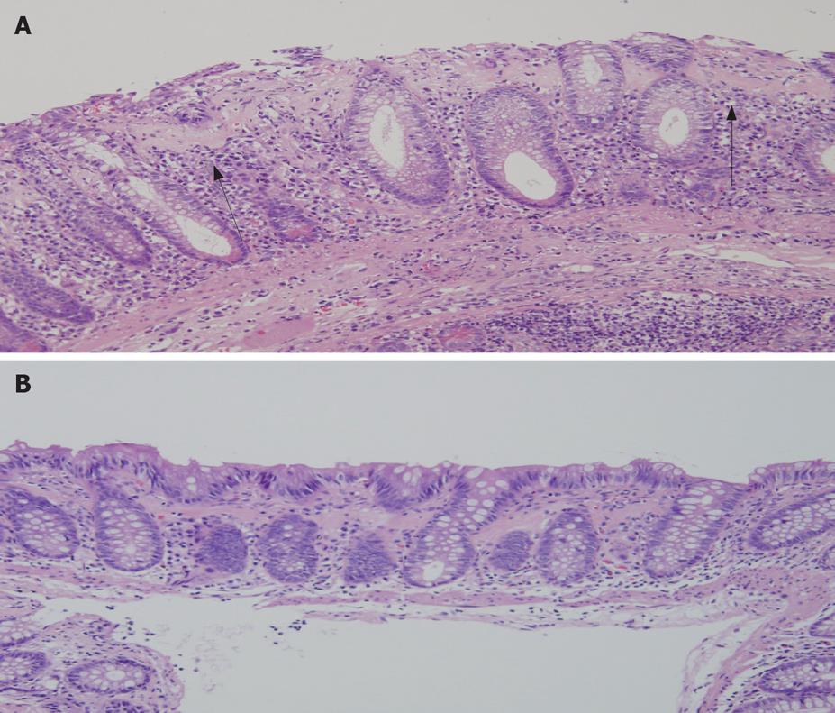
Figure 2 Histology of the biopsied specimen demonstrates subepithelial from eosinophilic band-like deposit (arrows), with increased lymphocytes and plasma cells.
Sloughing of surface epithelium is also shown (A). Epithelial detachment and inflammatory cells decreased, although the collagen band beneath the mucosa was not reduced (B).
- Citation: Sano S, Yamagami K, Tanaka A, Nishio M, Nakamura T, Kubo Y, Inoue T, Ueda W, Okawa K, Yoshioka K. A unique case of collagenous colitis presenting as protein-losing enteropathy successfully treated with prednisolone. World J Gastroenterol 2008; 14(39): 6083-6086
- URL: https://www.wjgnet.com/1007-9327/full/v14/i39/6083.htm
- DOI: https://dx.doi.org/10.3748/wjg.14.6083


