Copyright
©2008 The WJG Press and Baishideng.
World J Gastroenterol. Sep 21, 2008; 14(35): 5403-5411
Published online Sep 21, 2008. doi: 10.3748/wjg.14.5403
Published online Sep 21, 2008. doi: 10.3748/wjg.14.5403
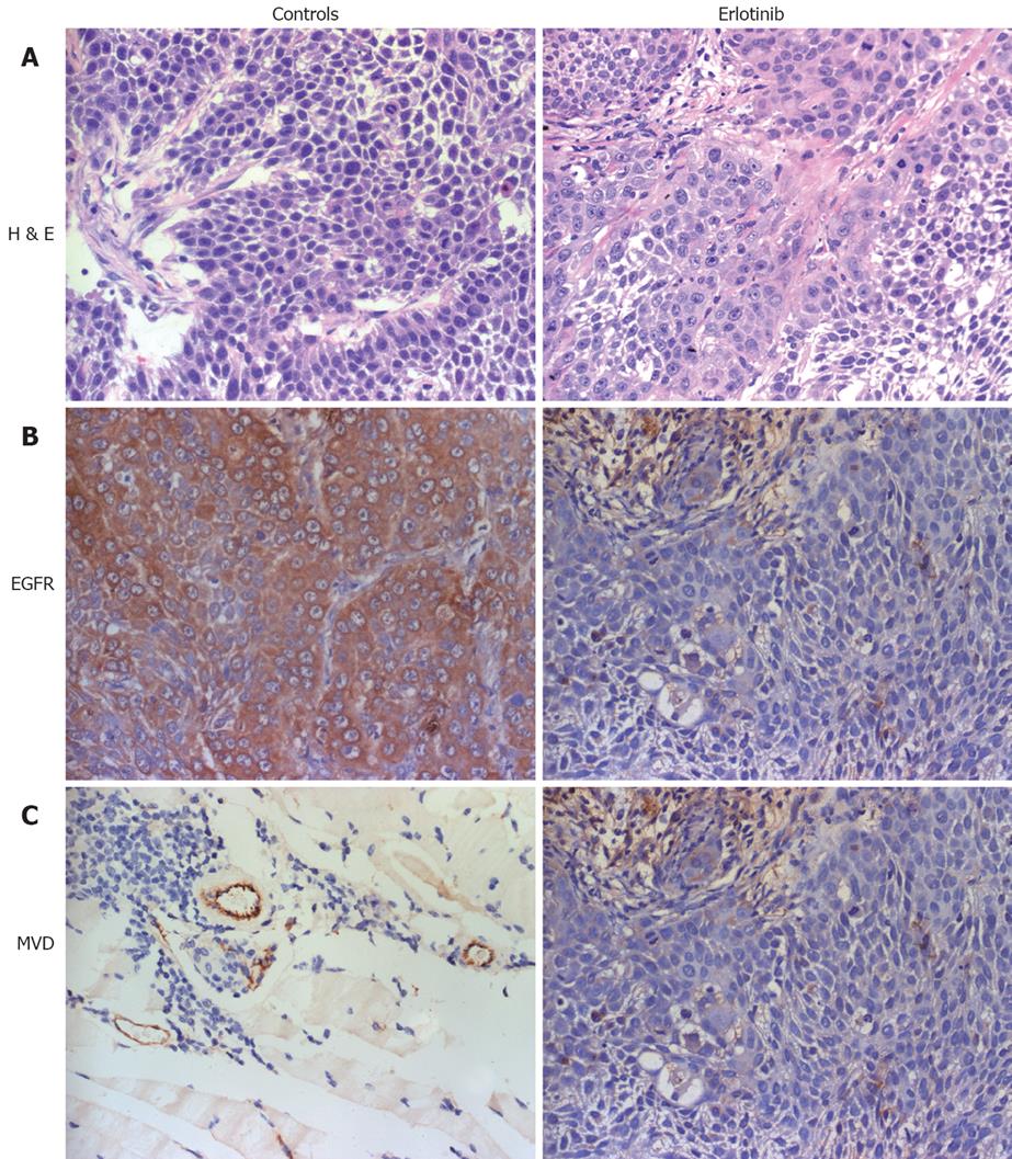
Figure 5 Expression of EGFR and the blood vessel endothelial cells in different treatment group in BxPC-3 mouse xenograft tissues.
IHC was used to determine expression levels of EGFR and evaluate tumor microvessel density. A: HE staining for each sample (× 400); B: Expression of EGFR in treatment group was decreased compared with the control (× 400); C: Microvessel density of erlotinib treated group was lower than that of the control group.
- Citation: Lu YY, Jing DD, Xu M, Wu K, Wang XP. Anti-tumor activity of erlotinib in the BxPC-3 pancreatic cancer cell line. World J Gastroenterol 2008; 14(35): 5403-5411
- URL: https://www.wjgnet.com/1007-9327/full/v14/i35/5403.htm
- DOI: https://dx.doi.org/10.3748/wjg.14.5403


