Copyright
©2008 The WJG Press and Baishideng.
World J Gastroenterol. Aug 21, 2008; 14(31): 4964-4967
Published online Aug 21, 2008. doi: 10.3748/wjg.14.4964
Published online Aug 21, 2008. doi: 10.3748/wjg.14.4964
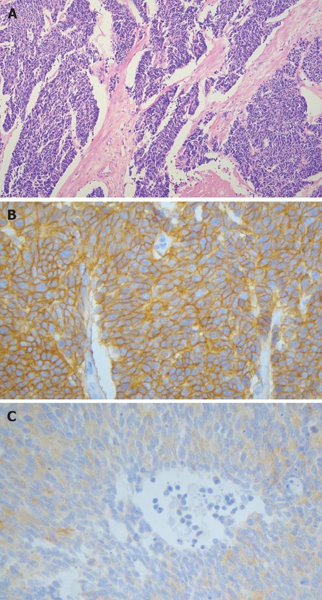
Figure 4 A: The tumor consisted of small round or oval cells with hyperchromatic nuclei and scant cytoplasm; B and C: Immunohistochemical staining for CD56 (B) and synaptophysin (C) reveals a positive reaction.
- Citation: Chung MS, Ha TK, Lee KG, Paik SS. A case of long survival in poorly differentiated small cell carcinoma of the pancreas. World J Gastroenterol 2008; 14(31): 4964-4967
- URL: https://www.wjgnet.com/1007-9327/full/v14/i31/4964.htm
- DOI: https://dx.doi.org/10.3748/wjg.14.4964


