Copyright
©2008 The WJG Press and Baishideng.
World J Gastroenterol. Aug 14, 2008; 14(30): 4763-4770
Published online Aug 14, 2008. doi: 10.3748/wjg.14.4763
Published online Aug 14, 2008. doi: 10.3748/wjg.14.4763
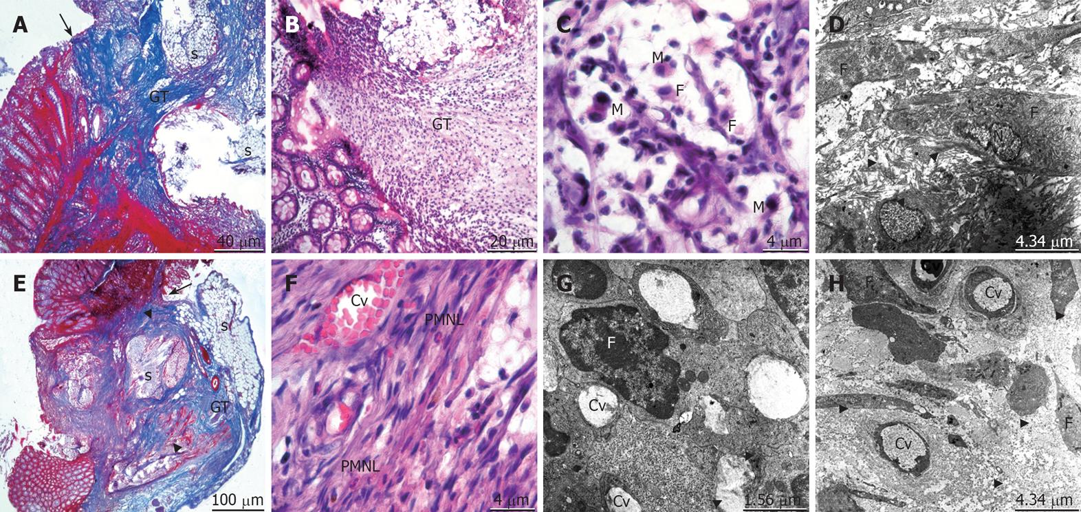
Figure 3 An overview of healing at day 7 after operation.
The micrographs “A, E” represent mallory-azan stained sections, “B, C, F” hematoxylin-eosin stained sections and “D, G, H” T.E.M. images of control (upper panels) and propolis (bottom panels) group. Arrow: The anastomosis site; Arrowhead: Collagen; GT: Granulation tissue; s: Suture material; M: Macrophage; F: Fibroblast; N: Nucleus; Cv: Capillary vessel.
- Citation: Kilicoglu SS, Kilicoglu B, Erdemli E. Ultrastructural view of colon anastomosis under propolis effect by transmission electron microscopy. World J Gastroenterol 2008; 14(30): 4763-4770
- URL: https://www.wjgnet.com/1007-9327/full/v14/i30/4763.htm
- DOI: https://dx.doi.org/10.3748/wjg.14.4763


