Copyright
©2008 The WJG Press and Baishidengs.
World J Gastroenterol. Aug 7, 2008; 14(29): 4694-4696
Published online Aug 7, 2008. doi: 10.3748/wjg.14.4694
Published online Aug 7, 2008. doi: 10.3748/wjg.14.4694
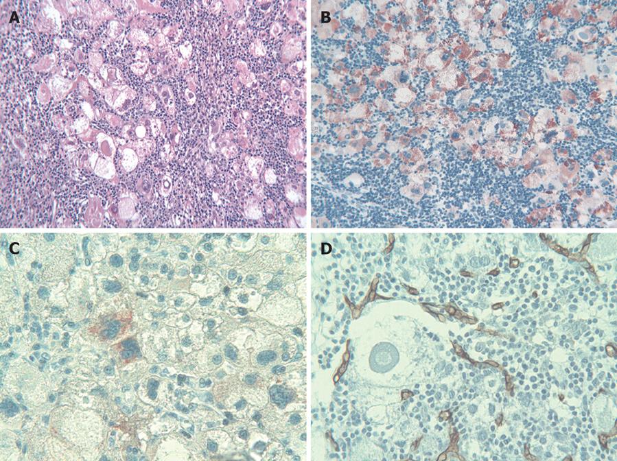
Figure 1 Histological picture characterized by a strong lymphoid intratumoral infiltrate (HE, × 250) (A), vast majority of neoplastic cells showing intense granular cytoplasmic reactivity for Hep Par 1 (× 250) (B), scattered atypical tumor cells showing cytoplasmic immunoreactivity for Glypican 3 (× 400) (C), and a large atypical tumor cell surrounded by CD34-positive newly formed capillaries (× 400) (D).
- Citation: Nemolato S, Fanni D, Naccarato AG, Ravarino A, Bevilacqua G, Faa G. Lymphoepithelioma-like hepatocellular carcinoma: A case report and a review of the literature. World J Gastroenterol 2008; 14(29): 4694-4696
- URL: https://www.wjgnet.com/1007-9327/full/v14/i29/4694.htm
- DOI: https://dx.doi.org/10.3748/wjg.14.4694


