Copyright
©2008 The WJG Press and Baishideng.
World J Gastroenterol. Jul 7, 2008; 14(25): 4093-4095
Published online Jul 7, 2008. doi: 10.3748/wjg.14.4093
Published online Jul 7, 2008. doi: 10.3748/wjg.14.4093
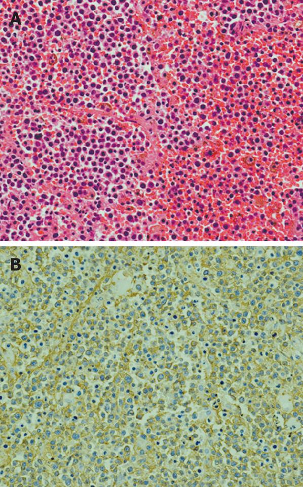
Figure 3 Photomicrographs of the tumor.
A: The tumor consists of diffuse sheets of large malignant lymphoid cells (HE, × 20); B: Immunostaining of the tumor. The tumor cells are positive for CD20 ( immunostaining, × 20).
- Citation: Hashimoto M, Umekita N, Noda K. Non-Hodgkin lymphoma as a cause of obstructive jaundice with simultaneous extrahepatic portal vein obstruction: A case report. World J Gastroenterol 2008; 14(25): 4093-4095
- URL: https://www.wjgnet.com/1007-9327/full/v14/i25/4093.htm
- DOI: https://dx.doi.org/10.3748/wjg.14.4093


