Copyright
©2008 The WJG Press and Baishideng.
World J Gastroenterol. Jun 7, 2008; 14(21): 3328-3337
Published online Jun 7, 2008. doi: 10.3748/wjg.14.3328
Published online Jun 7, 2008. doi: 10.3748/wjg.14.3328
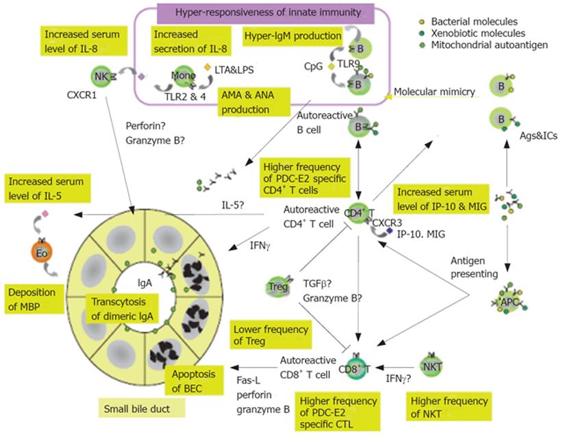
Figure 1 Model of pathogenic mechanisms in primary biliary cirrhosis (PBC).
PBC is initiated by an autoantigenic stimulus (upper, right) provided either by a bacterial mimic of the autoepitope of PDC-E2, a xenobiotically modified PDC-E2, or “spillage” of native mitochondrial autoantigens derived perhaps from apoptotic cells. Hyper-responsiveness of innate immunity (top, centre) can facilitate autoantigenicity; bacterial Cpg enhances IgM production and cellular expression of TLR9. Genetic susceptibility is critical overall, and depends particularly on multiple inherited deficits in immune tolerance, mostly as yet undefined. APCs that become activated (lower, right) by stimulation through TLRs present immunogenic self peptides (or mimics) via MHC Class II molecules to autoreactive CD4+ T lymphocytes (centre) which in turn activate CD8+ cytotoxic T lymphocytes and B lymphocytes that produce AMA. Treg lymphocytes (lower, centre) that normally restrain activated autoreactive T cells are deficient in PBC, thus further impeding T cell tolerance. Effector mechanisms converge on the target cell in PBC, the BEC (lower left), which can be damaged by injurious cytokines (IFN-γ) from CD4+ T cells, direct cytotoxicity (Fas-L, perforin, granzyme B) from CD8+ T cells, or transcytosis of IgA-AMA. A toxic effect might even be supplied by activated eosinophils (centre, left) by release of eosinophil MBP. BECs thus undergo apoptosis and in doing so contribute immunogenic mitochondrial PDC-E2 autoantigen to sustain a self-perpetuating autoimmunization process and, by reason of a BEC-specific anomaly of apoptosis retain PBC-E2 intact in apoptotic blebs (see text), so conferring particular vulnerability on these cells. Ags: Antigens; AMA: Antimitochondrial antibodies; ANA: Antinuclear antibodies; APC: Antigen-presenting cell; BEC: Biliary epithelial cells; CTL: Cytotoxic T lymphocytes; ICs: Immune complexes; IL: Interleukin; IFN: Interferon; IP-10: Interferon-γ-inducible protein 10; LTA: Lipoteichoic acid; LPS: Lipopolysaccharide; MIG: Monokine induced by γ-interferon; MBP: Major basic protein (primary cytotoxic granule protein); NKT: Natural killer T cells; PDC-E2: Pyruvate dehydrogenase complex E2; TGF: Transforming growth factor; TLR: Toll-like receptor; Treg: Regulatory T cells.
- Citation: Lleo A, Invernizzi P, Mackay IR, Prince H, Zhong RQ, Gershwin ME. Etiopathogenesis of primary biliary cirrhosis. World J Gastroenterol 2008; 14(21): 3328-3337
- URL: https://www.wjgnet.com/1007-9327/full/v14/i21/3328.htm
- DOI: https://dx.doi.org/10.3748/wjg.14.3328


