Copyright
©2008 The WJG Press and Baishideng.
World J Gastroenterol. May 14, 2008; 14(18): 2832-2837
Published online May 14, 2008. doi: 10.3748/wjg.14.2832
Published online May 14, 2008. doi: 10.3748/wjg.14.2832
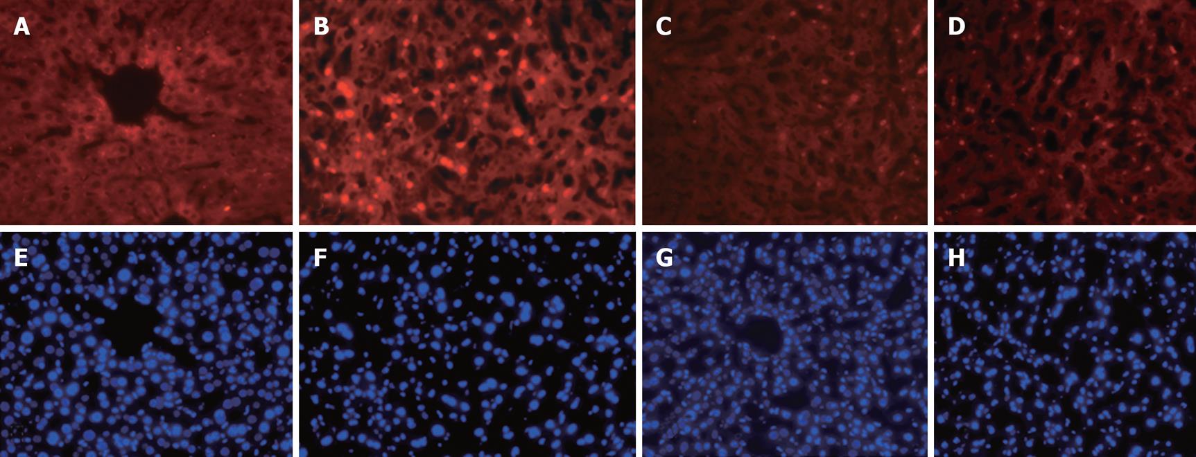
Figure 2 Representative micrographs of in situ TUNEL staining of apoptotic cells in liver tissue after the hepatic ischemia/reperfusion procedure.
A-D: The TUNEL staining assay was performed as described in the text, and the positive apoptotic cells were stained in red (× 100). A: Control; B: I/R procedure plus N.S. (6 h after starting reperfusion); C: I/R plus apocynin; D: I/R plus allopurinol. E-H: The corresponding liver sections stained with DAPI to illustrate nuclei as a cell number control.
- Citation: Liu PG, He SQ, Zhang YH, Wu J. Protective effects of apocynin and allopurinol on ischemia/reperfusion-induced liver injury in mice. World J Gastroenterol 2008; 14(18): 2832-2837
- URL: https://www.wjgnet.com/1007-9327/full/v14/i18/2832.htm
- DOI: https://dx.doi.org/10.3748/wjg.14.2832


