Copyright
©2008 The WJG Press and Baishideng.
World J Gastroenterol. May 7, 2008; 14(17): 2776-2779
Published online May 7, 2008. doi: 10.3748/wjg.14.2776
Published online May 7, 2008. doi: 10.3748/wjg.14.2776
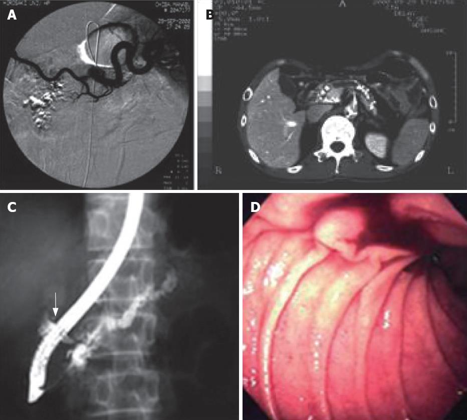
Figure 3 Case 2.
Pseudoaneurysm in pancreatic pseudocyst. A: Angiography failed to detect a bleeding point; B: CT scan shows multiple calcifications at the whole pancreas and dilatation of main pancreatic duct; C: Endoscopic retrograde pancreatography displays a dilatated branch of pancreatic duct at head of pancreas (arrow); D: Endoscopy reveals bleeding from papilla of Vater.
- Citation: Toyoki Y, Hakamada K, Narumi S, Nara M, Ishido K, Sasaki M. Hemosuccus pancreaticus: Problems and pitfalls in diagnosis and treatment. World J Gastroenterol 2008; 14(17): 2776-2779
- URL: https://www.wjgnet.com/1007-9327/full/v14/i17/2776.htm
- DOI: https://dx.doi.org/10.3748/wjg.14.2776


