Copyright
©2008 The WJG Press and Baishideng.
World J Gastroenterol. Apr 14, 2008; 14(14): 2272-2276
Published online Apr 14, 2008. doi: 10.3748/wjg.14.2272
Published online Apr 14, 2008. doi: 10.3748/wjg.14.2272
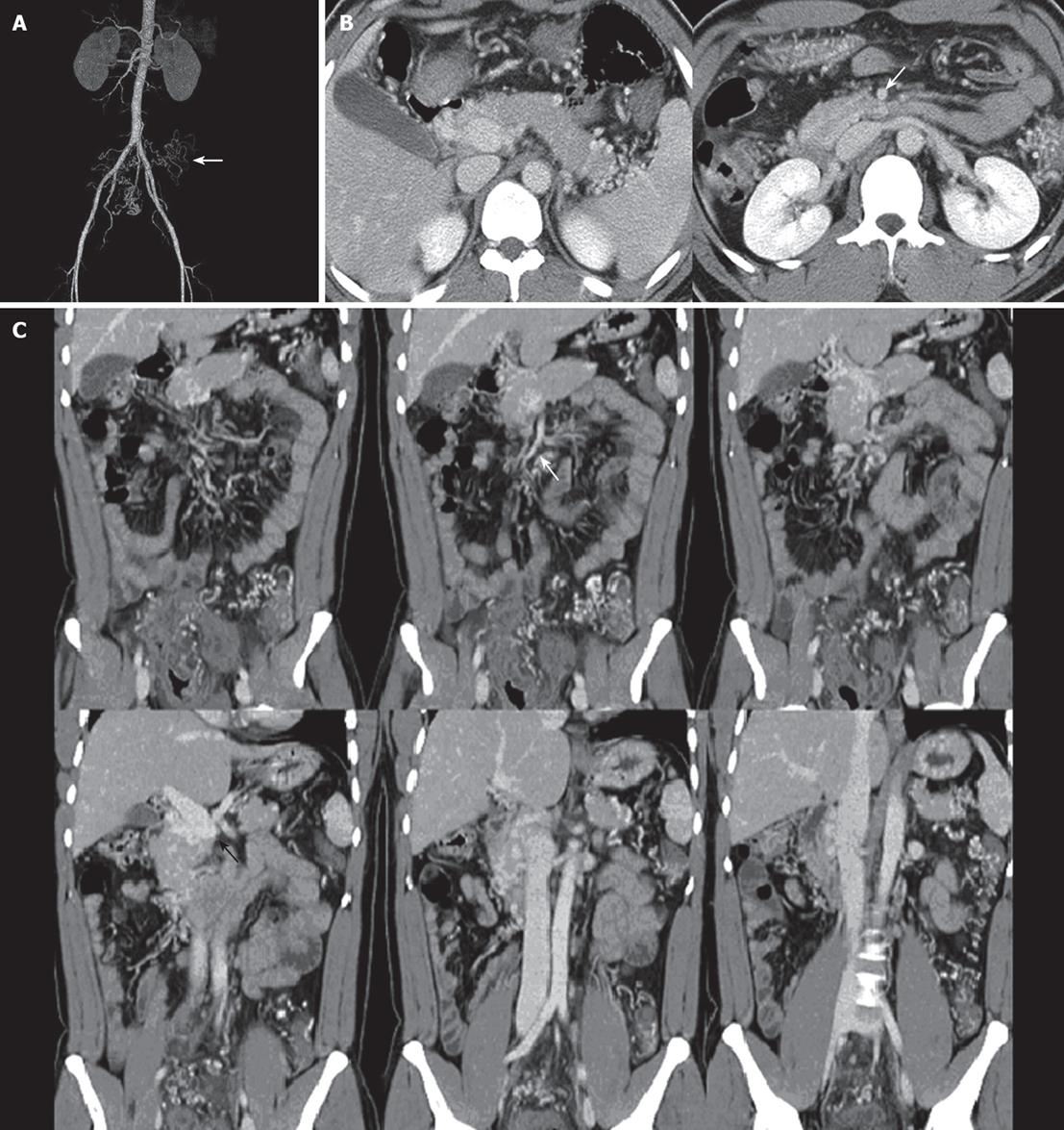
Figure 3 Abdominal CT arteriography.
A: Normal shape and course of SMA, IMA, and splenic artery, tortuous and dilated distal branches of SMA and IMA around rectosigmoid colon; B: An axial and C: Coronal reformatted abdominal CT reveal the absent splenic vein, SMV, and IMV with numerous intra- and peripancreatic collaterals. Distal end portal vein is abruptly ended (black arrow) and SMA is normal (white arrow). Note prominent dilated pericolic arteries and venous collaterals, and markedly
thickened sigmoid colon.
- Citation: Hwang SS, Chung WC, Lee KM, Kim HJ, Paik CN, Yang JM. Ischemic colitis due to obstruction of mesenteric and splenic veins: A case report. World J Gastroenterol 2008; 14(14): 2272-2276
- URL: https://www.wjgnet.com/1007-9327/full/v14/i14/2272.htm
- DOI: https://dx.doi.org/10.3748/wjg.14.2272


