Copyright
©2008 The WJG Press and Baishideng.
World J Gastroenterol. Apr 14, 2008; 14(14): 2272-2276
Published online Apr 14, 2008. doi: 10.3748/wjg.14.2272
Published online Apr 14, 2008. doi: 10.3748/wjg.14.2272
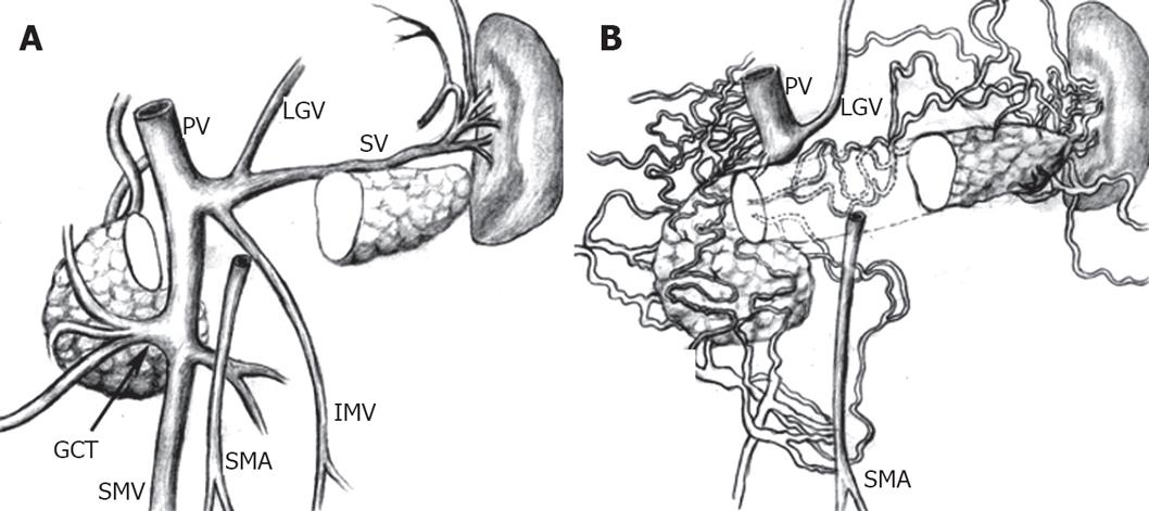
Figure 2 Dynamic abdominal CT.
A: The normal anatomy of the main portal vein and its tributaries including the portal vein (PV), left gastric vein (LGV), gastrocolic trunk (GCT), splenic vein (SV), SMA, SMV, and IMV are schematically illustrated; B: The normal mesenteric vein (SMV and IMV) and splenic vein are absent. Numerous collateral venous channels developed to drain venous blood from gastrointestinal tract to portal vein.
- Citation: Hwang SS, Chung WC, Lee KM, Kim HJ, Paik CN, Yang JM. Ischemic colitis due to obstruction of mesenteric and splenic veins: A case report. World J Gastroenterol 2008; 14(14): 2272-2276
- URL: https://www.wjgnet.com/1007-9327/full/v14/i14/2272.htm
- DOI: https://dx.doi.org/10.3748/wjg.14.2272


