
















©2007 Baishideng Publishing Group Co.
World J Gastroenterol. Mar 7, 2007; 13(9): 1408-1421
Published online Mar 7, 2007. doi: 10.3748/wjg.v13.i9.1408
Published online Mar 7, 2007. doi: 10.3748/wjg.v13.i9.1408
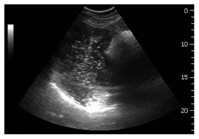
Figure 3 An oblique frontal section applied for ultrasound scanning of the proximal stomach.
The top margin of the fundus is shown as a white line in the bottom of the image. A proximal gastric diameter is outlined in the frontal section by tracing normally to the longitudinal axis of the proximal stomach, thus indicating the size of the proximal stomach.
- Citation: Gilja OH, Hatlebakk JG, Ødegaard S, Berstad A, Viola I, Giertsen C, Hausken T, Gregersen H. Advanced imaging and visualization in gastrointestinal disorders. World J Gastroenterol 2007; 13(9): 1408-1421
- URL: https://www.wjgnet.com/1007-9327/full/v13/i9/1408.htm
- DOI: https://dx.doi.org/10.3748/wjg.v13.i9.1408