Copyright
©2007 Baishideng Publishing Group Co.
World J Gastroenterol. Mar 7, 2007; 13(9): 1378-1383
Published online Mar 7, 2007. doi: 10.3748/wjg.v13.i9.1378
Published online Mar 7, 2007. doi: 10.3748/wjg.v13.i9.1378
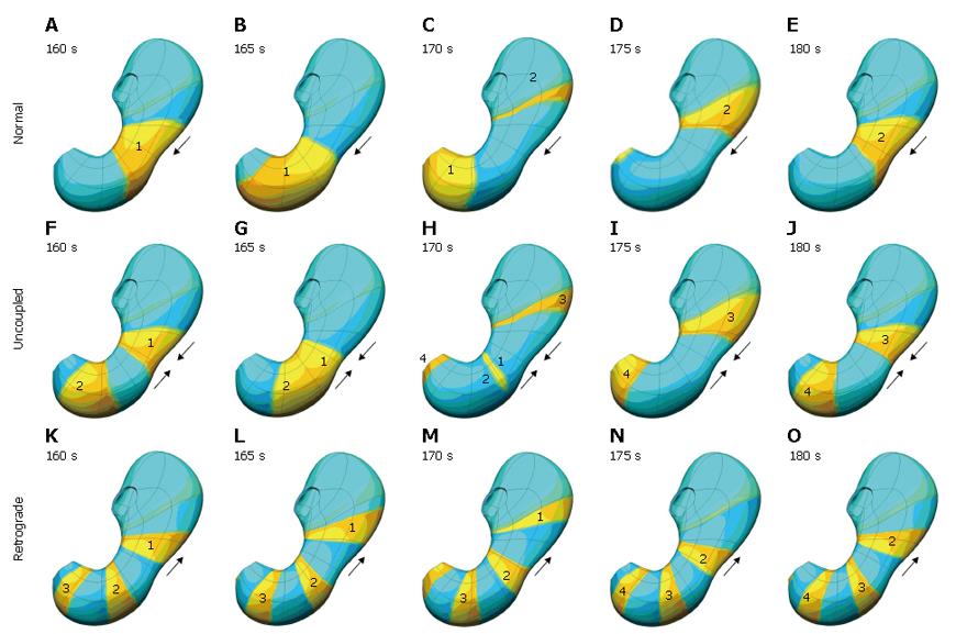
Figure 6 Top row (A-E) shows normal slow wave activity at 5 second interval.
In this case the antrum is entrained by the slow wave activity originating in the corpus, is shown. In the second row (F-J), the corpus and antrum maintain the same frequency of slow wave activity, but in this situation, the antrum is not being entrained by the activity of the corpus. The bottom row (K-O) illustrates what happens when the intrinsic frequency of the antrum exceeds that of the corpus, resulting in some retrograde slow wave behaviour.
- Citation: Cheng LK, Komuro R, Austin TM, Buist ML, Pullan AJ. Anatomically realistic multiscale models of normal and abnormal gastrointestinal electrical activity. World J Gastroenterol 2007; 13(9): 1378-1383
- URL: https://www.wjgnet.com/1007-9327/full/v13/i9/1378.htm
- DOI: https://dx.doi.org/10.3748/wjg.v13.i9.1378


