Copyright
©2007 Baishideng Publishing Group Co.
World J Gastroenterol. Feb 28, 2007; 13(8): 1275-1278
Published online Feb 28, 2007. doi: 10.3748/wjg.v13.i8.1275
Published online Feb 28, 2007. doi: 10.3748/wjg.v13.i8.1275
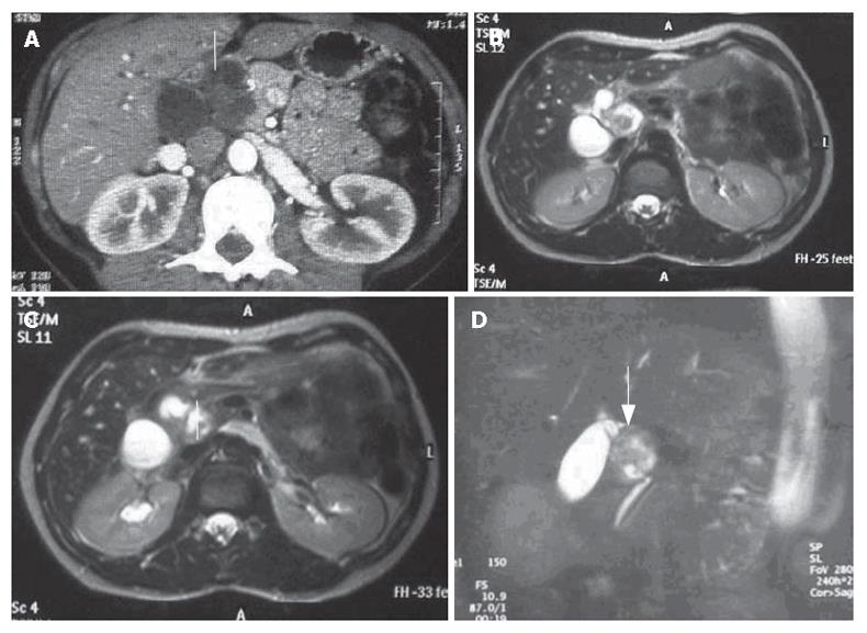
Figure 1 A: CT scan showing a very heterogeneous formation with a liquid content in close contiguity to the CBD (white arrow); B, C, D: Cholangio-MRI reveals a solid formation with a hypointensive signal in the weighted T1 sequences at the pancreatic isthmus (white arrows) with dilation of the upstream bile ducts.
- Citation: Fenoglio L, Severini S, Cena P, Migliore E, Bracco C, Pomero F, Panzone S, Cavallero GB, Silvestri A, Brizio R, Borghi F. Common bile duct schwannoma: A case report and review of literature. World J Gastroenterol 2007; 13(8): 1275-1278
- URL: https://www.wjgnet.com/1007-9327/full/v13/i8/1275.htm
- DOI: https://dx.doi.org/10.3748/wjg.v13.i8.1275


