Copyright
©2007 Baishideng Publishing Group Co.
World J Gastroenterol. Feb 28, 2007; 13(8): 1257-1261
Published online Feb 28, 2007. doi: 10.3748/wjg.v13.i8.1257
Published online Feb 28, 2007. doi: 10.3748/wjg.v13.i8.1257
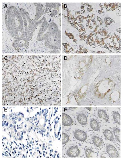
Figure 1 Typical immunohistochemical staining for Eag1 in colorectal cancer and adenoma.
A, B, C: Positive staining of colorectal cancers; D: Positive staining of metastasis tissue from greater omentum; E: Negative control of colorectal cancer; F: Positive staining from one case of colonic adenoma.
- Citation: Ding XW, Yan JJ, An P, Lü P, Luo HS. Aberrant expression of ether à go-go potassium channel in colorectal cancer patients and cell lines. World J Gastroenterol 2007; 13(8): 1257-1261
- URL: https://www.wjgnet.com/1007-9327/full/v13/i8/1257.htm
- DOI: https://dx.doi.org/10.3748/wjg.v13.i8.1257


