Copyright
©2007 Baishideng Publishing Group Co.
World J Gastroenterol. Feb 7, 2007; 13(5): 809-812
Published online Feb 7, 2007. doi: 10.3748/wjg.v13.i5.809
Published online Feb 7, 2007. doi: 10.3748/wjg.v13.i5.809
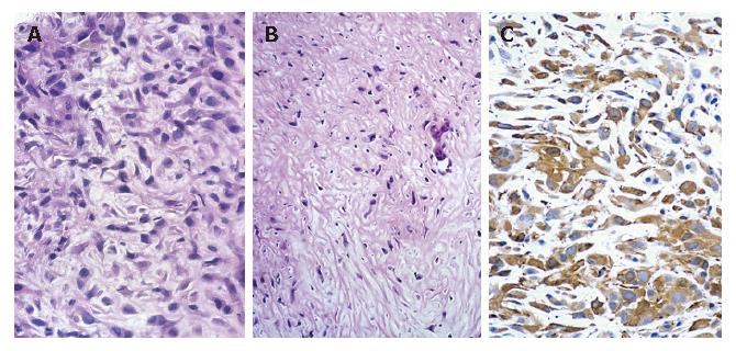
Figure 2 Needle biopsy showing a sarcomatous area consisting of interlacing bundles of atypical spindle cells [hematoxylin-eosin stain; magnification x 200 (A), x 100 (B)] and immunohistochemical staining showing positive α-SMA (C).
- Citation: Sumiyoshi S, Kikuyama M, Matsubayashi Y, Kageyama F, Ide Y, Kobayashi Y, Nakamura H. Carcinosarcoma of the liver with mesenchymal differentiation. World J Gastroenterol 2007; 13(5): 809-812
- URL: https://www.wjgnet.com/1007-9327/full/v13/i5/809.htm
- DOI: https://dx.doi.org/10.3748/wjg.v13.i5.809


