Copyright
©2007 Baishideng Publishing Group Co.
World J Gastroenterol. Dec 28, 2007; 13(48): 6512-6517
Published online Dec 28, 2007. doi: 10.3748/wjg.v13.i48.6512
Published online Dec 28, 2007. doi: 10.3748/wjg.v13.i48.6512
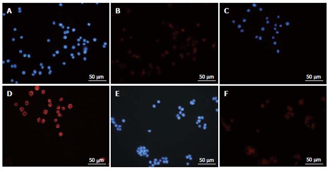
Figure 6 Immunofluorescence staining with Atg8 (LC3).
SW480 (A), COLO (B) and WiDr (C) cells were cultured with MK615 at 300 μg/mL for 6 h. Nuclei were stained with DAPI (A, C and E), and Atg8 (LC3) was localized in the cytoplasm (B, D and F; note the central blank area in the cells).
-
Citation: Mori S, Sawada T, Okada T, Ohsawa T, Adachi M, Keiichi K. New anti-proliferative agent, MK615, from Japanese apricot “
Prunus mume ” induces striking autophagy in colon cancer cellsin vitro . World J Gastroenterol 2007; 13(48): 6512-6517 - URL: https://www.wjgnet.com/1007-9327/full/v13/i48/6512.htm
- DOI: https://dx.doi.org/10.3748/wjg.v13.i48.6512


