Copyright
©2007 Baishideng Publishing Group Inc.
World J Gastroenterol. Dec 14, 2007; 13(46): 6269-6273
Published online Dec 14, 2007. doi: 10.3748/wjg.v13.i46.6269
Published online Dec 14, 2007. doi: 10.3748/wjg.v13.i46.6269
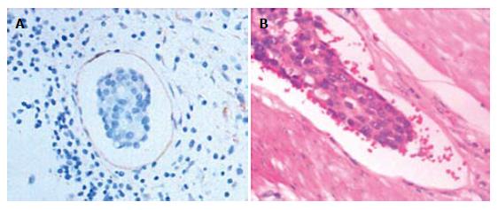
Figure 1 Immunohistochemical staining (A) and hematoxylin-eosin staining (B) of tumor cells (× 400) showing a tumor cell cluster in vascular spaces with brown-stained endothelial cells and tumor cells in blood vessel spaces with erythrocytes surrounded.
- Citation: Wang YD, Wu P, Mao JD, Huang H, Zhang F. Relationship between vascular invasion and microvessel density and micrometastasis. World J Gastroenterol 2007; 13(46): 6269-6273
- URL: https://www.wjgnet.com/1007-9327/full/v13/i46/6269.htm
- DOI: https://dx.doi.org/10.3748/wjg.v13.i46.6269


