Copyright
©2007 Baishideng Publishing Group Inc.
World J Gastroenterol. Oct 21, 2007; 13(39): 5196-5207
Published online Oct 21, 2007. doi: 10.3748/wjg.v13.i39.5196
Published online Oct 21, 2007. doi: 10.3748/wjg.v13.i39.5196
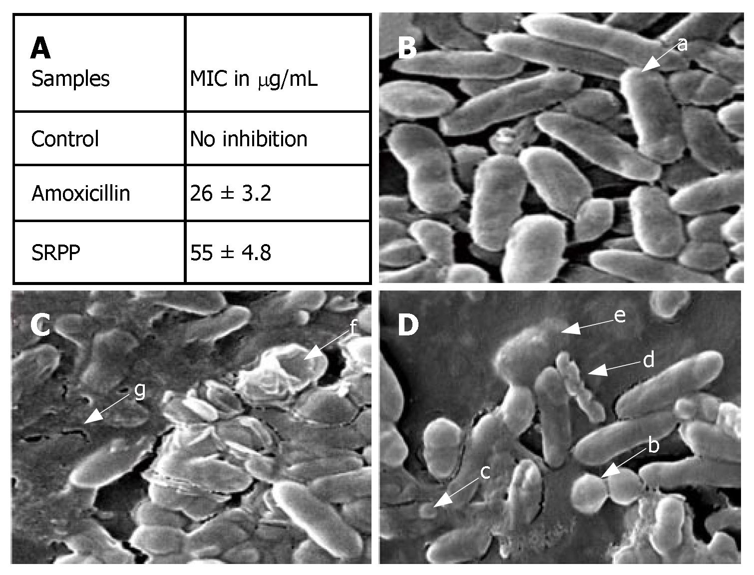
Figure 5 Effect of SRPP on H pylori; Minimum Inhibitory Concentration (MIC) (A) was established by serial dilution technique; B-D indicate the scanning electron microscopic pictures at 15 k magnification of control (B), SRPP (C) and amoxicillin (D) treated H pylori.
Untreated control cultures indicate uniform rod shaped (a) H pylori cells. Amoxicillin treatment showed coccoid form (b), blebbing (c), fragmented (d) and lysed (e) cells. SRPP treatment in addition indicates cavity formation (f) with disrupted structures (g).
-
Citation: Srikanta B, Siddaraju M, Dharmesh S. A novel phenol-bound pectic polysaccharide from
Decalepis hamiltonii with multi-step ulcer preventive activity. World J Gastroenterol 2007; 13(39): 5196-5207 - URL: https://www.wjgnet.com/1007-9327/full/v13/i39/5196.htm
- DOI: https://dx.doi.org/10.3748/wjg.v13.i39.5196


