Copyright
©2007 Baishideng Publishing Group Co.
World J Gastroenterol. Sep 28, 2007; 13(36): 4897-4902
Published online Sep 28, 2007. doi: 10.3748/wjg.v13.i36.4897
Published online Sep 28, 2007. doi: 10.3748/wjg.v13.i36.4897
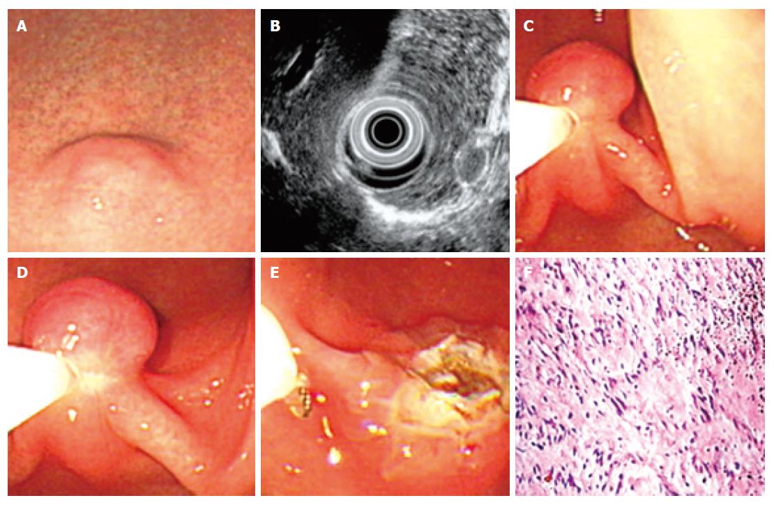
Figure 2 Endoscopic views for “pushing” resection of a leiomyoma.
A: A sessile leiomyoma at antrum of stomach; B: EUS revealed that the mass originated from muscularis mucosa; C: The leiomyoma was pushed by cannula to form a semipedunculation and then captured by snare; D: The captured leiomyoma was resected by high-frequency electrosurgical current; E: The endoscopic view for the cauterization burn of leiomyoma after resection; F: The histologic view of leiomyoma after resection (HE, x 200 ).
- Citation: Zhou XD, Lv NH, Chen HX, Wang CW, Zhu X, Xu P, Chen YX. Endoscopic management of gastrointestinal smooth muscle tumor. World J Gastroenterol 2007; 13(36): 4897-4902
- URL: https://www.wjgnet.com/1007-9327/full/v13/i36/4897.htm
- DOI: https://dx.doi.org/10.3748/wjg.v13.i36.4897


