Copyright
©2007 Baishideng Publishing Group Co.
World J Gastroenterol. Sep 28, 2007; 13(36): 4881-4890
Published online Sep 28, 2007. doi: 10.3748/wjg.v13.i36.4881
Published online Sep 28, 2007. doi: 10.3748/wjg.v13.i36.4881
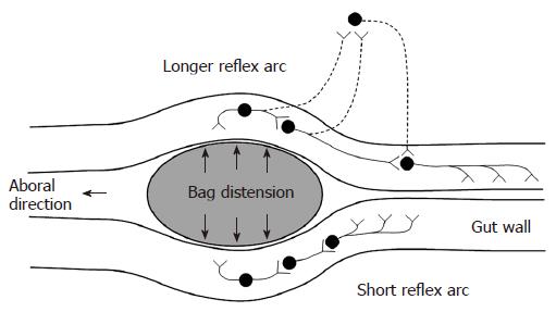
Figure 2 Schematic illustration of the neural pathways involved in distension-induced contraction.
The primary afferent neurons, the excitatory interneurons and efferent neurons are shown. The dotted neurons illustrate extrinsic pathways. The induction of contraction on the bag depends on the local reflex arc (bottom). The measurement 6 cm proximal to the bag depends on a longer reflex arc including extrinsic pathways (top).
- Citation: Frøkjær JB, Andersen SD, Ejskjær N, Funch-Jensen P, Drewes AM, Gregersen H. Impaired contractility and remodeling of the upper gastrointestinal tract in diabetes mellitus type-1. World J Gastroenterol 2007; 13(36): 4881-4890
- URL: https://www.wjgnet.com/1007-9327/full/v13/i36/4881.htm
- DOI: https://dx.doi.org/10.3748/wjg.v13.i36.4881


