Copyright
©2007 Baishideng Publishing Group Co.
World J Gastroenterol. Aug 21, 2007; 13(31): 4242-4248
Published online Aug 21, 2007. doi: 10.3748/wjg.v13.i31.4242
Published online Aug 21, 2007. doi: 10.3748/wjg.v13.i31.4242
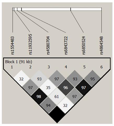
Figure 1 Linkage disequilibrium plot across the CLOCK gene.
The horizontal white line depicts the 117-kb DNA segment of chromosome 4q12 analyzed in our sample. The 6 tagSNPs locations are indicated by hatch marks. A linkage disequilibrium plot is depicted in the bottom part of the figure. Each diamond represents the magnitude of LD for a single pair of markers, with colors indicating strong LD (black, r2 = 1.0) and no LD (white, r2 = 0) as the extremes (different gray tones indicate intermediate LD). Numbers inside the diamonds stand for D’ values.
-
Citation: Sookoian S, Castaño G, Gemma C, Gianotti TF, Pirola CJ. Common genetic variations in
CLOCK transcription factor are associated with nonalcoholic fatty liver disease. World J Gastroenterol 2007; 13(31): 4242-4248 - URL: https://www.wjgnet.com/1007-9327/full/v13/i31/4242.htm
- DOI: https://dx.doi.org/10.3748/wjg.v13.i31.4242


