Copyright
©2007 Baishideng Publishing Group Inc.
World J Gastroenterol. Jul 7, 2007; 13(25): 3472-3477
Published online Jul 7, 2007. doi: 10.3748/wjg.v13.i25.3472
Published online Jul 7, 2007. doi: 10.3748/wjg.v13.i25.3472
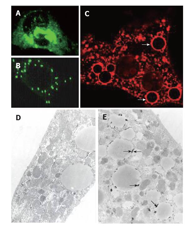
Figure 2 A and B: Immunofluorescent stain for transfected HeLa cells and primary hepatocytes (C) (× 60).
Two cytoplasmic distribution patterns of the core protein in HeLa cells were obtained-condensed perinuclear (A) and disseminated granular (B) patterns. The core protein was present exclusively in the cytoplasm of hepatocytes and some of it exhibited the vesicular pattern (C, × 60, arrows). Under the electron microscope (D and E, × 100), the vesicular structures were found to be lipid vesicles and the core protein was located just underneath the surface of the vesicles (E, arrows), whereas the cells transfected with empty plasmid (PUGH16-3) showed no core associated with the lipid vesicles (D).
-
Citation: Chang ML, Chen JC, Yeh CT, Sheen IS, Tai DI, Chang MY, Chiu CT, Lin DY, Bissell DM. Topological and evolutional relationships between HCV core protein and hepatic lipid vesicles: Studies
in vitro and in conditionally transgenic mice. World J Gastroenterol 2007; 13(25): 3472-3477 - URL: https://www.wjgnet.com/1007-9327/full/v13/i25/3472.htm
- DOI: https://dx.doi.org/10.3748/wjg.v13.i25.3472


