
















©2007 Baishideng Publishing Group Co.
World J Gastroenterol. Jun 21, 2007; 13(23): 3255-3258
Published online Jun 21, 2007. doi: 10.3748/wjg.v13.i23.3255
Published online Jun 21, 2007. doi: 10.3748/wjg.v13.i23.3255
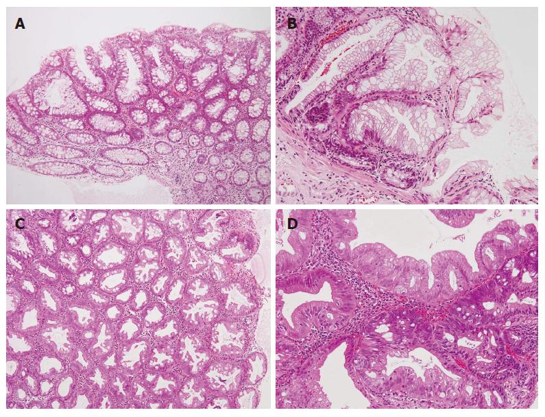
Figure 2 A: Tubular adenoma located in the transverse colon (HE, × 100).
The lesion shows duct proliferation and cells that have mild nuclear atypia; B: Hyperplastic polyp located in the transverse colon (HE, × 200). Serrated ducts with no atypical features are observed; C: Hyperplastic polyp located in the ascending colon (HE, × 100). Serrated ducts with no atypical features are observed. The inflammatory infiltration is mainly composed of lymphocytes in the lamina propria; D: High-grade serrated adenoma located in the sigmoid colon (HE, × 200). Papillary or serrated structures with obvious atypical cells are seen. Atypical structures, such as fusion of the tubular structure, are also shown. There is no evidence of malignancy.
- Citation: Kurobe M, Abe K, Kinoshita N, Anami M, Tokai H, Ryu Y, Wen CY, Kanematsu T, Hayashi T. Hyperplastic polyposis associated with two asynchronous colon cancers. World J Gastroenterol 2007; 13(23): 3255-3258
- URL: https://www.wjgnet.com/1007-9327/full/v13/i23/3255.htm
- DOI: https://dx.doi.org/10.3748/wjg.v13.i23.3255