
















©2007 Baishideng Publishing Group Co.
World J Gastroenterol. Jun 14, 2007; 13(22): 3080-3083
Published online Jun 14, 2007. doi: 10.3748/wjg.v13.i22.3080
Published online Jun 14, 2007. doi: 10.3748/wjg.v13.i22.3080
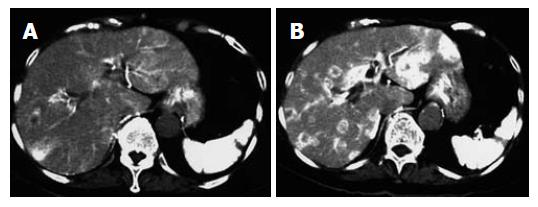
Figure 1 A 78 years old woman with liver metastasis from pancreatic cancer.
A:Conventional angiographic CT; B: Pharmacoangiographic CT with angiotensin II. The tumor to liver contrast is increased, and well demarcated low density area in the liver is now highly indicative of liver metastasis.
- Citation: Ishikawa T, Ushiki T, Kamimura H, Togashi T, Tsuchiya A, Watanabe K, Seki KI, Ohta H, Yoshida T, Takeda K, Kamimura T. Angiotensin-II administration is useful for the detection of liver metastasis from pancreatic cancer during pharmacoangiographic computed tomography. World J Gastroenterol 2007; 13(22): 3080-3083
- URL: https://www.wjgnet.com/1007-9327/full/v13/i22/3080.htm
- DOI: https://dx.doi.org/10.3748/wjg.v13.i22.3080