Copyright
©2007 Baishideng Publishing Group Co.
World J Gastroenterol. Jun 14, 2007; 13(22): 3063-3070
Published online Jun 14, 2007. doi: 10.3748/wjg.v13.i22.3063
Published online Jun 14, 2007. doi: 10.3748/wjg.v13.i22.3063
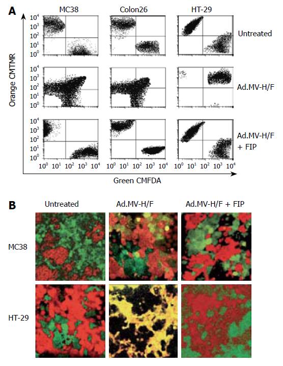
Figure 1 Quantification of cell-cell fusion by flow cytometry and laser scanning confocal microscopy.
A: Twenty-four hours later cells were analyzed for dye colocalization by flow cytometry. As a control we used the synthetic fusion inhibitory peptide (FIP); B: In addition we analyzed the cells 36 h after transduction with Ad.MV-H/F by confocal laser scanning microscopy. Transduction of Colon26 cells with Ad.MV-H/F resulted in similar cell-cell fusion as in MC38 cells (data not shown). One representative experiment out of three is shown.
-
Citation: Hoffmann D, Bayer W, Wildner O.
In situ tumor vaccination with adenovirus vectors encoding measles virus fusogenic membrane proteins and cytokines. World J Gastroenterol 2007; 13(22): 3063-3070 - URL: https://www.wjgnet.com/1007-9327/full/v13/i22/3063.htm
- DOI: https://dx.doi.org/10.3748/wjg.v13.i22.3063


