
















©2007 Baishideng Publishing Group Co.
World J Gastroenterol. Jun 14, 2007; 13(22): 3043-3046
Published online Jun 14, 2007. doi: 10.3748/wjg.v13.i22.3043
Published online Jun 14, 2007. doi: 10.3748/wjg.v13.i22.3043
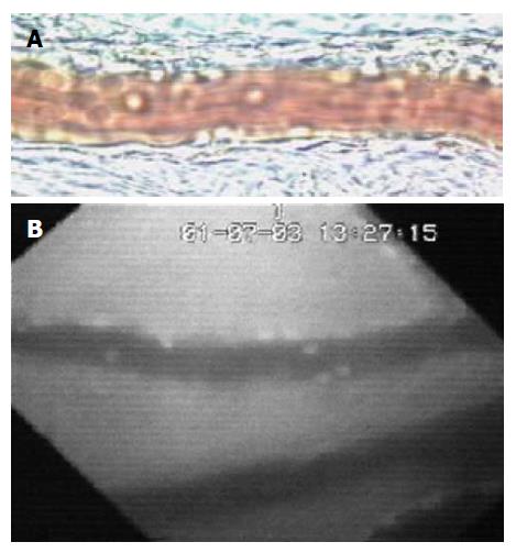
Figure 2 Image of mesenteric (A) and small bowel (B) venules was obtained by Intravital microscopic.
Rolling and firm adherent leukocytes can be easily identified by transillumination (A) or fluorescent staining (B).
- Citation: Mollà M, Panés J. Radiation-induced intestinal inflammation. World J Gastroenterol 2007; 13(22): 3043-3046
- URL: https://www.wjgnet.com/1007-9327/full/v13/i22/3043.htm
- DOI: https://dx.doi.org/10.3748/wjg.v13.i22.3043