Copyright
©2007 Baishideng Publishing Group Co.
World J Gastroenterol. May 28, 2007; 13(20): 2841-2845
Published online May 28, 2007. doi: 10.3748/wjg.v13.i20.2841
Published online May 28, 2007. doi: 10.3748/wjg.v13.i20.2841
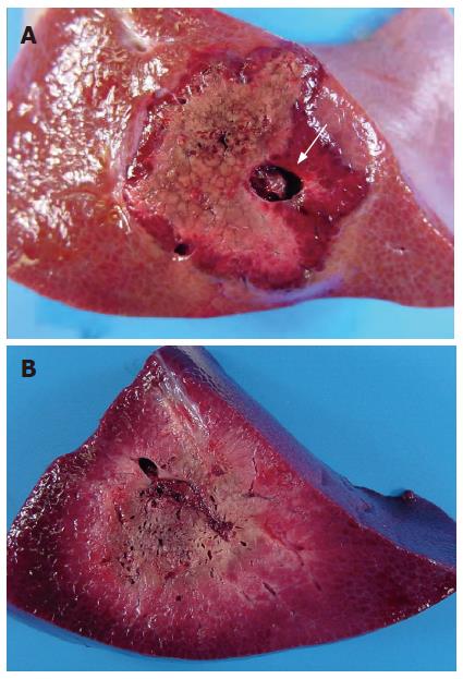
Figure 4 Region of necrosis avoiding large vessels (white arrow) in group with RFA alone (A), and showing ill-defined boundaries of coagulation necrosis and increase of the necrotic area in the group with RFA following LP-TAE (B).
- Citation: Nakai M, Sato M, Sahara S, Kawai N, Tanihata H, Kimura M, Terada M. Radiofrequency ablation in a porcine liver model: Effects of transcatheter arterial embolization with iodized oil on ablation time, maximum output, and coagulation diameter as well as angiographic characteristics. World J Gastroenterol 2007; 13(20): 2841-2845
- URL: https://www.wjgnet.com/1007-9327/full/v13/i20/2841.htm
- DOI: https://dx.doi.org/10.3748/wjg.v13.i20.2841


