Copyright
©2007 Baishideng Publishing Group Co.
World J Gastroenterol. Jan 7, 2007; 13(1): 91-103
Published online Jan 7, 2007. doi: 10.3748/wjg.v13.i1.91
Published online Jan 7, 2007. doi: 10.3748/wjg.v13.i1.91
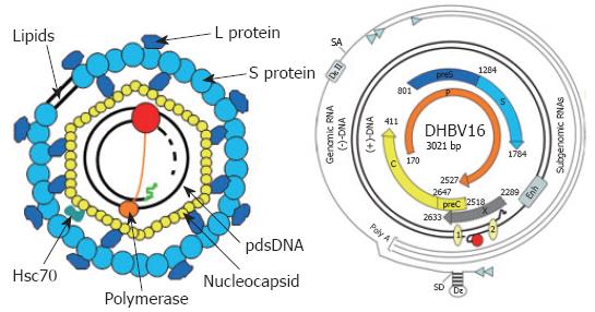
Figure 2 Virion structure and genome organization of avian hepadnaviruses.
The viral envelope is derived from hepatocellular membranes and contains the viral surface proteins S and L. It covers the nucleocapsid harbouring the viral genome with the covalently linked terminal protein domain (TP, orange circle) of the polymerase (P, red circle) and cellular proteins like Hsc70. The genome is organized as depicted. The various transcripts are indicated by thin lines with the small arrowhead indicating the start sites. The partially double stranded, viral DNA with the covalently bound TP domain of P (red circle) is symbolized by the thicker lines. The numbered circles 1 and 2 on the viral DNA represent the direct repeats (DR). Enh represents the enhancer domain. The ORFs encoding core (C), polymerase (P), and the surface proteins (preS and S) are symbolized by thick arrows. Epsilon (ξ) is the stem loop structure on the pgRNA which acts as an encapsidation signal and replication origin. The second encapsidation element DξII is unique to avian hepatitis B viruses, since the mammalian counterparts lack this RNA structure. SD and SA represent the major splice donor and acceptor sites, respectively.
- Citation: Funk A, Mhamdi M, Will H, Sirma H. Avian hepatitis B viruses: Molecular and cellular biology, phylogenesis, and host tropism. World J Gastroenterol 2007; 13(1): 91-103
- URL: https://www.wjgnet.com/1007-9327/full/v13/i1/91.htm
- DOI: https://dx.doi.org/10.3748/wjg.v13.i1.91


