
















©2007 Baishideng Publishing Group Co.
World J Gastroenterol. Jan 7, 2007; 13(1): 48-64
Published online Jan 7, 2007. doi: 10.3748/wjg.v13.i1.48
Published online Jan 7, 2007. doi: 10.3748/wjg.v13.i1.48
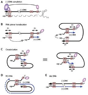
Figure 8 RC-DNA formation.
A: (-)-DNA completion. The DNA primer, still linked to TP, is extended from DR1* to the 5´ end of pgRNA. The RNA is simultaneously degraded by the RH domain, except for its capped 5´ terminal region including 5´ DR1; the fate of the poly-adenylated 3´ end is unclear; B: RNA primer translocation (second template switch). The RNA primer translocates to DR2, and is extended to the 5´ end of (-)-DNA. 3´ r and 5´ r denote an about 10 nt redundancy on the (-)-DNA. As above, several cis-elements appear to promote close proximity of the DR1 donor and the DR2 acceptor, as schematically indicated in the right hand figure; C: Circularization (third template switch). Having copied 5´ r, the growing 3´ end of the (+)-DNA switches to 3´ r on the (-)-DNA, enabling further elongation. This reaction must involve juxtaposition of 5´ r and 3´ r. For easier comprehension, the switch is also depicted on the basis of the representation shown on the right of Figure 8B; both are topologically equivalent; D: RC-DNA. Extension on the (-)-DNA template creates a set of (+)-DNA strands of various length; E: Double-stranded linear (dsL) DNA. This minor DNA form originates when the RNA primer, having failed to translocate to DR2, is extended from its original position ("in situ priming").
- Citation: Beck J, Nassal M. Hepatitis B virus replication. World J Gastroenterol 2007; 13(1): 48-64
- URL: https://www.wjgnet.com/1007-9327/full/v13/i1/48.htm
- DOI: https://dx.doi.org/10.3748/wjg.v13.i1.48