Copyright
©2007 Baishideng Publishing Group Co.
World J Gastroenterol. Jan 7, 2007; 13(1): 22-38
Published online Jan 7, 2007. doi: 10.3748/wjg.v13.i1.22
Published online Jan 7, 2007. doi: 10.3748/wjg.v13.i1.22
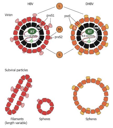
Figure 1 Schematic presentation of human (HBV) and duck (DHBV) hepatitis B virus.
The viral DNA is drawn as a single or double line. The viral polymerase is depicted with the primer domain (pr) and the reverse transcription domain (RT). The nucleocapsid (core or HBc/DHBc) is shown in black. Reported encapsidated cellular proteins are omitted. For HBV the surface proteins L, M and S are shown with the S-domain, the preS2-domain and the preS1-domain, whereas for DHBV, L- and S-surface proteins with preS and S-domain are depicted. St, truncated form of DHBV S-surface protein. Non-infectious subviral particles of HBV are shown in filamentous and spherical form and in larger spheroids in the case of DHBV.
- Citation: Glebe D, Urban S. Viral and cellular determinants involved in hepadnaviral entry. World J Gastroenterol 2007; 13(1): 22-38
- URL: https://www.wjgnet.com/1007-9327/full/v13/i1/22.htm
- DOI: https://dx.doi.org/10.3748/wjg.v13.i1.22


