Copyright
©2006 Baishideng Publishing Group Co.
World J Gastroenterol. Feb 21, 2006; 12(7): 1092-1097
Published online Feb 21, 2006. doi: 10.3748/wjg.v12.i7.1092
Published online Feb 21, 2006. doi: 10.3748/wjg.v12.i7.1092
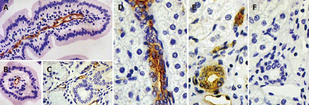
Figure 4 Immunolocalization of pAQP1 in pig gastrointestinal tract and exocrine glands.
Specific pAQP1 labeling of central lacteals was seen by immunoperoxidase staining of longitudal (A) and traverse (B) sections of the small intestinal villi using an affinity-purified AQP1 antibody. Arrows indicate the endothelium of central lacteals; A salivary gland section showing specific labeling of microvessel endothelium indicated by arrows (C); Specific pAQP1 labeling was seen in the epithelium of intrahepatic bile ducts in longitudal (D) and traverse (E) sections. Arrows indicate heavy pAQP1 staining in the apical domain of bile duct epithelial cells. Arowhead indicates pAQP1 labeling in the endothelium of periductal microvessels; A consecutive section of E showing immunostaining with AQP1 antibody preabsorbed with the immunizing peptide (F).
- Citation: Jin SY, Liu YL, Xu LN, Jiang Y, Wang Y, Yang BX, Yang H, Ma TH. Cloning and characterization of porcine aquaporin 1 water channel expressed extensively in gastrointestinal system. World J Gastroenterol 2006; 12(7): 1092-1097
- URL: https://www.wjgnet.com/1007-9327/full/v12/i7/1092.htm
- DOI: https://dx.doi.org/10.3748/wjg.v12.i7.1092


