Copyright
©2006 Baishideng Publishing Group Co.
World J Gastroenterol. Feb 14, 2006; 12(6): 990-992
Published online Feb 14, 2006. doi: 10.3748/wjg.v12.i6.990
Published online Feb 14, 2006. doi: 10.3748/wjg.v12.i6.990
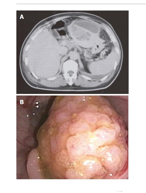
Figure 1 Case 1.
A: Enhancement abdominal computed tomography (CT) showed a 4-cm liver abscess in left lobe of the liver; B: Colonofibroscopy showed a 3-cm pedunculated polyp in the sigmoid colon.
- Citation: Lai HC, Chan CY, Peng CY, Chen CB, Huang WH. Pyogenic liver abscess associated with large colonic tubulovillous adenoma. World J Gastroenterol 2006; 12(6): 990-992
- URL: https://www.wjgnet.com/1007-9327/full/v12/i6/990.htm
- DOI: https://dx.doi.org/10.3748/wjg.v12.i6.990


