Copyright
©2006 Baishideng Publishing Group Co.
World J Gastroenterol. Feb 14, 2006; 12(6): 858-867
Published online Feb 14, 2006. doi: 10.3748/wjg.v12.i6.858
Published online Feb 14, 2006. doi: 10.3748/wjg.v12.i6.858
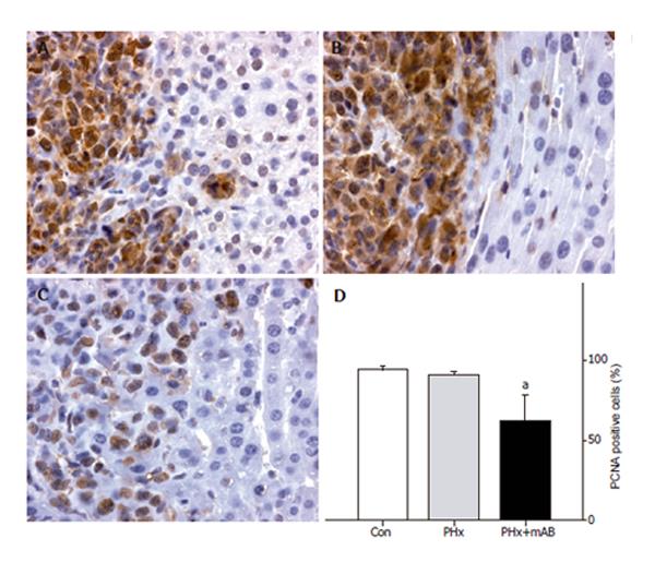
Figure 7 PCNA immunohistochemistry and quantitative analysis of the number of PCNA-positive cells (given in percent of all cells) in liver tumors of control mice (A, Con), after hepatectomy (B, PHx), and after hepatectomy and additional anti-MIP-2 treatment (C, PHx+mAB).
Tumor cells display massive PCNA staining, in particular to those located within the tumor margin. By this, these positive cells sharply demarcate the tumor from the surrounding liver tissue (A, B, and C). Quantitative analysis revealed that neutralization of MIP-2 significantly reduces the number of PCNA-positive tumor cells (D). Mean ± SE; aP < 0.05 vs Con. Magnifications (A-C) ×175.
- Citation: Kollmar O, Menger MD, Schilling MK. Macrophage inflammatory protein-2 contributes to liver resection-induced acceleration of hepatic metastatic tumor growth. World J Gastroenterol 2006; 12(6): 858-867
- URL: https://www.wjgnet.com/1007-9327/full/v12/i6/858.htm
- DOI: https://dx.doi.org/10.3748/wjg.v12.i6.858


