Copyright
©2006 Baishideng Publishing Group Co.
World J Gastroenterol. Dec 28, 2006; 12(48): 7869-7873
Published online Dec 28, 2006. doi: 10.3748/wjg.v12.i48.7869
Published online Dec 28, 2006. doi: 10.3748/wjg.v12.i48.7869
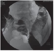
Figure 1 Solitary mass type: Enhanced CT showing multiple enlarged lymph nodes fused into uneven density, huge lobular tu-mors with abdo-minal aorta and inferior vena cava encased and ascites.
- Citation: Yu RS, Zhang WM, Liu YQ. CT diagnosis of 52 patients with lymphoma in abdominal lymph nodes. World J Gastroenterol 2006; 12(48): 7869-7873
- URL: https://www.wjgnet.com/1007-9327/full/v12/i48/7869.htm
- DOI: https://dx.doi.org/10.3748/wjg.v12.i48.7869


