Copyright
©2006 Baishideng Publishing Group Co.
World J Gastroenterol. Dec 21, 2006; 12(47): 7717-7719
Published online Dec 21, 2006. doi: 10.3748/wjg.v12.i47.7717
Published online Dec 21, 2006. doi: 10.3748/wjg.v12.i47.7717
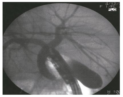
Figure 2 Endoscopic retrograde cholangiography of the patient.
A stone was extracted from the main duct. The gallbladder can be observed on the left side.
- Citation: Aydin U, Unalp O, Yazici P, Gurcu B, Sozbilen M, Coker A. Laparoscopic cholecystectomy in a patient with situs inversus totalis. World J Gastroenterol 2006; 12(47): 7717-7719
- URL: https://www.wjgnet.com/1007-9327/full/v12/i47/7717.htm
- DOI: https://dx.doi.org/10.3748/wjg.v12.i47.7717


