Copyright
©2006 Baishideng Publishing Group Co.
World J Gastroenterol. Oct 28, 2006; 12(40): 6556-6558
Published online Oct 28, 2006. doi: 10.3748/wjg.v12.i40.6556
Published online Oct 28, 2006. doi: 10.3748/wjg.v12.i40.6556
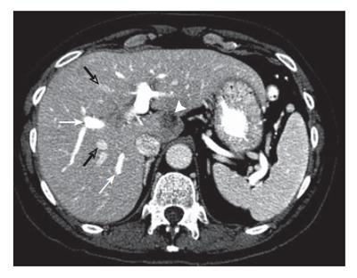
Figure 1 Contrast enhanced CT at the level of the portal bifurcation before treatment shows metastasis in segment I (arrowhead), opacified right anterior and posterior segmental portal veins (white arrows), and opacified middle and right hepatic veins (open arrows).
- Citation: Donadon M, Vauthey JN, Loyer EM, Charnsangavej C, Abdalla EK. Portal thrombosis and steatosis after preoperativechemotherapy with FOLFIRI-bevacizumab for colorectal liver metastases. World J Gastroenterol 2006; 12(40): 6556-6558
- URL: https://www.wjgnet.com/1007-9327/full/v12/i40/6556.htm
- DOI: https://dx.doi.org/10.3748/wjg.v12.i40.6556


