Copyright
©2006 Baishideng Publishing Group Co.
World J Gastroenterol. Oct 28, 2006; 12(40): 6531-6535
Published online Oct 28, 2006. doi: 10.3748/wjg.v12.i40.6531
Published online Oct 28, 2006. doi: 10.3748/wjg.v12.i40.6531
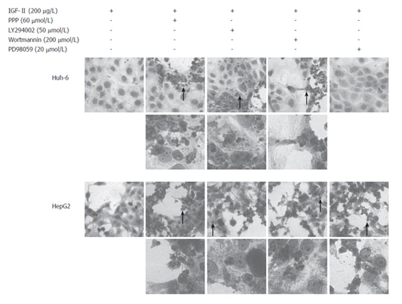
Figure 5 Huh-6 and HepG2 dead due to apoptosis.
HE staining was performed to analyze morphological changes after addition of inhibitors. Many Huh-6 cells were dead treated with PPP (60 μmol/L), LY294002 (50 μmol/L), or Wortmannin (200 μmol/L) while HepG2 with PPP, LY294002, Wortmannin, or PD98059 (20 μmol/L). Most of the dead cells had pyknotic or fragmented nuclei (arrows), suggesting apoptosis. Areas indicated by arrows were magnified (x 400).
- Citation: Tomizawa M, Saisho H. Signaling pathway of insulin-like growth factor-II as a target of molecular therapy for hepatoblastoma. World J Gastroenterol 2006; 12(40): 6531-6535
- URL: https://www.wjgnet.com/1007-9327/full/v12/i40/6531.htm
- DOI: https://dx.doi.org/10.3748/wjg.v12.i40.6531


