Copyright
©2006 Baishideng Publishing Group Co.
World J Gastroenterol. Oct 28, 2006; 12(40): 6531-6535
Published online Oct 28, 2006. doi: 10.3748/wjg.v12.i40.6531
Published online Oct 28, 2006. doi: 10.3748/wjg.v12.i40.6531
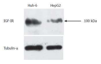
Figure 2 Western blot analysis clearly shows specific bands to IGF-IR.
Protein was isolated 72 h after stimulation with IGF-II (200 μg/L). The same membrane was reprobed with anti-Tubulin-α antibody to confirm an equal amount of protein loadings.
- Citation: Tomizawa M, Saisho H. Signaling pathway of insulin-like growth factor-II as a target of molecular therapy for hepatoblastoma. World J Gastroenterol 2006; 12(40): 6531-6535
- URL: https://www.wjgnet.com/1007-9327/full/v12/i40/6531.htm
- DOI: https://dx.doi.org/10.3748/wjg.v12.i40.6531


