Copyright
©2006 Baishideng Publishing Group Co.
World J Gastroenterol. Sep 28, 2006; 12(36): 5834-5845
Published online Sep 28, 2006. doi: 10.3748/wjg.v12.i36.5834
Published online Sep 28, 2006. doi: 10.3748/wjg.v12.i36.5834
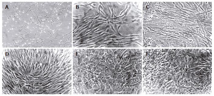
Figure 4 Morphology of human mesenchymal stem cells from bone marrow and adipose tissue during the differentiation protocol.
Cells were induced to differentiate by using a sequential addition of growth factors, cytokines and hormones. Morphology of passage 2 BMSC (A) and ADSC (B) cells. No significant morphological changes were observed in BMSC (C) nor in ADSC (D) cells during the step-1 differentiation. However, both BMSC (E) and ADSC (F) cells significantly changed the morphology, and developed a polygonal shape during step-2 differentiation (magnification 20 x for all pictures).
- Citation: Taléns-Visconti R, Bonora A, Jover R, Mirabet V, Carbonell F, Castell JV, Gómez-Lechón MJ. Hepatogenic differentiation of human mesenchymal stem cells from adipose tissue in comparison with bone marrow mesenchymal stem cells. World J Gastroenterol 2006; 12(36): 5834-5845
- URL: https://www.wjgnet.com/1007-9327/full/v12/i36/5834.htm
- DOI: https://dx.doi.org/10.3748/wjg.v12.i36.5834


