Copyright
©2006 Baishideng Publishing Group Co.
World J Gastroenterol. Sep 21, 2006; 12(35): 5735-5738
Published online Sep 21, 2006. doi: 10.3748/wjg.v12.i35.5735
Published online Sep 21, 2006. doi: 10.3748/wjg.v12.i35.5735
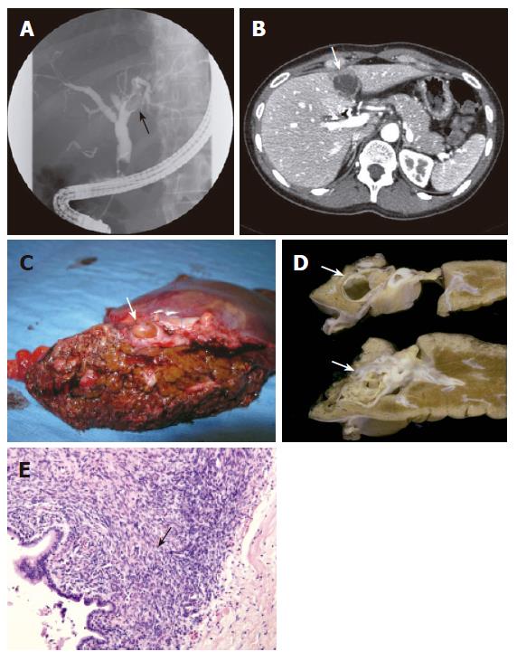
Figure 2 ERCP showing a filling defect due to an intraluminal lesion (arrow) in the left hepatic duct (A), CT-scan showing a cystic lesion with internal septations measuring 3.
6 cm located in segment 4 (arrow) (B), specimen after left hemihepatectomy showing macroscopic features of a large lesion (arrow) inside the left bile duct filling up the entire lumen (C), macroscopical cut sections of a multicystic lesion (arrows) encapsulated by a thick fibrous capsule arising from the left hepatic duct (D), microscopical features showing columnar mucinous epithelium with underlying dense-cellular stroma resembling ovarian stroma (arrow) (HE X 200) (E) in case 3.
- Citation: Erdogan D, Busch OR, Rauws EA, Delden OMV, Gouma DJ, Gulik TMV. Obstructive jaundice due to hepatobiliary cystadenoma or cystadenocarcinoma. World J Gastroenterol 2006; 12(35): 5735-5738
- URL: https://www.wjgnet.com/1007-9327/full/v12/i35/5735.htm
- DOI: https://dx.doi.org/10.3748/wjg.v12.i35.5735


