Copyright
©2006 Baishideng Publishing Group Co.
World J Gastroenterol. Sep 14, 2006; 12(34): 5573-5576
Published online Sep 14, 2006. doi: 10.3748/wjg.v12.i34.5573
Published online Sep 14, 2006. doi: 10.3748/wjg.v12.i34.5573
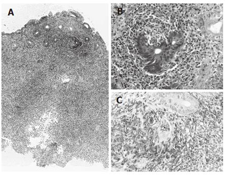
Figure 2 Biopsy specimens histologically showing diffuse proliferation of atypical small lymphocytes in the mucosal layer (A: x 40 magnification, HE) and glandular destruction (B: x 200 magnification, HE).
These lymphocytes immunohistochemically showing diffusely positive staining for L-26 (C; x 200 magnification, ABC method).
- Citation: Matsuo S, Mizuta Y, Hayashi T, Susumu S, Tsutsumi R, Azuma T, Yamaguchi S. Mucosa-associated lymphoid tissue lymphoma of the transverse colon: A case report. World J Gastroenterol 2006; 12(34): 5573-5576
- URL: https://www.wjgnet.com/1007-9327/full/v12/i34/5573.htm
- DOI: https://dx.doi.org/10.3748/wjg.v12.i34.5573


