Copyright
©2006 Baishideng Publishing Group Co.
World J Gastroenterol. Aug 28, 2006; 12(32): 5247-5252
Published online Aug 28, 2006. doi: 10.3748/wjg.v12.i32.5247
Published online Aug 28, 2006. doi: 10.3748/wjg.v12.i32.5247
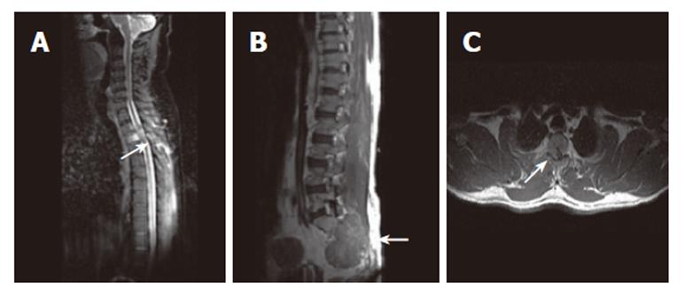
Figure 2 A: Sagittal MR image of cervicodorsal spine showing vertebral lesions with most prominent D3 vertebral lesion (arrow); B: Post contrast sagittal T1W SE MR image of the same patient showing lesion involving S2, S3 vertebrae (arrow) with associated large soft tissue component; C: T1W SE axial post contrast MR image showing right lateral epidural soft issue enhancing component (arrow) causing cord compression.
- Citation: Doval DC, Bhatia K, Vaid AK, Pavithran K, Sharma JB, Hazarika D, Jena A. Spinal cord compression secondary to bone metastases from hepatocellular carcinoma. World J Gastroenterol 2006; 12(32): 5247-5252
- URL: https://www.wjgnet.com/1007-9327/full/v12/i32/5247.htm
- DOI: https://dx.doi.org/10.3748/wjg.v12.i32.5247


