Copyright
©2006 Baishideng Publishing Group Co.
World J Gastroenterol. Aug 14, 2006; 12(30): 4859-4865
Published online Aug 14, 2006. doi: 10.3748/wjg.v12.i30.4859
Published online Aug 14, 2006. doi: 10.3748/wjg.v12.i30.4859
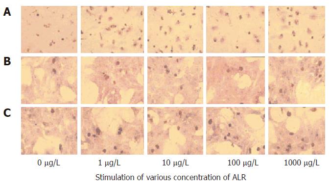
Figure 2 BrdU immunohistochemistry.
The nuclei of BrdU-positive cells were stained brown. A: Kupffer cells; B: Hepatocytes; C: Hepatocytes conditioned by Kupffer cells (× 100).
- Citation: Wang CP, Zhou L, Su SH, Chen Y, Lu YY, Wang F, Jia HJ, Feng YY, Yang YP. Augmenter of liver regeneration promotes hepatocyte proliferation induced by Kupffer cells. World J Gastroenterol 2006; 12(30): 4859-4865
- URL: https://www.wjgnet.com/1007-9327/full/v12/i30/4859.htm
- DOI: https://dx.doi.org/10.3748/wjg.v12.i30.4859


