Copyright
©2006 Baishideng Publishing Group Co.
World J Gastroenterol. Jul 28, 2006; 12(28): 4588-4592
Published online Jul 28, 2006. doi: 10.3748/wjg.v12.i28.4588
Published online Jul 28, 2006. doi: 10.3748/wjg.v12.i28.4588
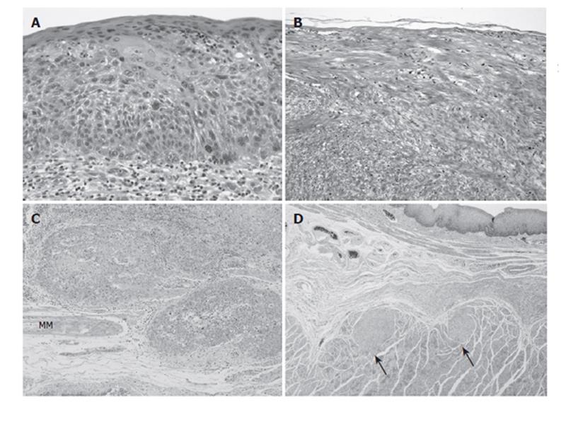
Figure 5 High-power view of the regions indicated by small letters in Figure 4.
A: Intraepithelial SCC; B: The SMT has low cellularity, appears strongly eosinophilic on hematoxylin-eosin staining, and is composed of interlaced smooth-muscle cells with hypovascularity and no mitosis; C: The SCC nest has invaded beyond the muscularis mucosae (MM); D: Two tiny leiomyomas, both originating from the muscular layer.
- Citation: Iwaya T, Maesawa C, Uesugi N, Kimura T, Ikeda K, Kimura Y, Mitomo S, Ishida K, Sato N, Wakabayashi G. Coexistence of esophageal superficial carcinoma and multiple leiomyomas: A case report. World J Gastroenterol 2006; 12(28): 4588-4592
- URL: https://www.wjgnet.com/1007-9327/full/v12/i28/4588.htm
- DOI: https://dx.doi.org/10.3748/wjg.v12.i28.4588


