Copyright
©2006 Baishideng Publishing Group Co.
World J Gastroenterol. Jul 28, 2006; 12(28): 4588-4592
Published online Jul 28, 2006. doi: 10.3748/wjg.v12.i28.4588
Published online Jul 28, 2006. doi: 10.3748/wjg.v12.i28.4588
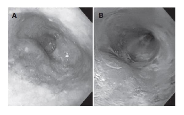
Figure 2 A: Endoscopic examination demonstrates a polypoid lesion with a partially irregular surface; B: Chromoendoscopy with Lugol’s iodine solution demonstrates a polypoid lesion located within the non-staining area and normal mucosa covering part of the surface.
- Citation: Iwaya T, Maesawa C, Uesugi N, Kimura T, Ikeda K, Kimura Y, Mitomo S, Ishida K, Sato N, Wakabayashi G. Coexistence of esophageal superficial carcinoma and multiple leiomyomas: A case report. World J Gastroenterol 2006; 12(28): 4588-4592
- URL: https://www.wjgnet.com/1007-9327/full/v12/i28/4588.htm
- DOI: https://dx.doi.org/10.3748/wjg.v12.i28.4588


