Copyright
©2006 Baishideng Publishing Group Co.
World J Gastroenterol. Jul 28, 2006; 12(28): 4517-4523
Published online Jul 28, 2006. doi: 10.3748/wjg.v12.i28.4517
Published online Jul 28, 2006. doi: 10.3748/wjg.v12.i28.4517
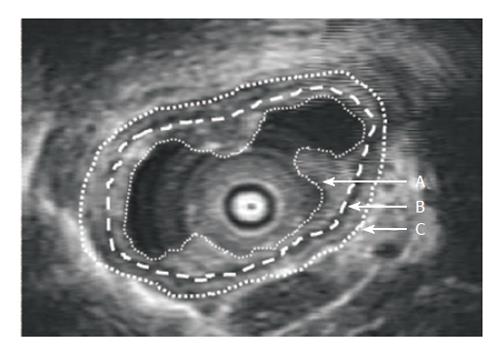
Figure 1 Transversal ultrasonographic image of the non-distended esophagus.
The stippled lines indicate manual tracings of the mucosal inner surface (A) and the inner (B) and outer (C) lining of the combined muscle layers.
- Citation: Larsen E, Reddy H, Drewes AM, Arendt-Nielsen L, Gregersen H. Ultrasonographic study of mechanosensory properties in human esophagus during mechanical distension. World J Gastroenterol 2006; 12(28): 4517-4523
- URL: https://www.wjgnet.com/1007-9327/full/v12/i28/4517.htm
- DOI: https://dx.doi.org/10.3748/wjg.v12.i28.4517


