Copyright
©2006 Baishideng Publishing Group Co.
World J Gastroenterol. Jul 14, 2006; 12(26): 4232-4236
Published online Jul 14, 2006. doi: 10.3748/wjg.v12.i26.4232
Published online Jul 14, 2006. doi: 10.3748/wjg.v12.i26.4232
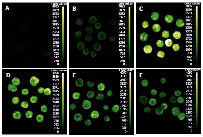
Figure 2 Tet prevented LPS-induced nuclear translocation of p65 in isolated pancreatic acinar cells.
P65 was visualized by indirect immunofluorescence staining using rabbit anti-p65 polyclonal antibodies (1:100) which only recognized NF-κB p65. Goat anti-rabbit antibodies (1:100) conjugated to FITC was performed, and visualized under a confocal microscope. Nuclear p65 was observed at 1 h (C), 4 h (D) after treatment with LPS (10 mg/L) but not in saline-treated controls at 1 h (B), nor in negative control (A). Nuclear localization of p65 and the fluorescence in cytoplasm were markedly reduced by Tet 100 μmol/L at 1 (E), 4 h (F), compared with that of LPS group at the same time (Original magnification × 400).
- Citation: Zhang H, Li YY, Wu XZ. Effect of Tetrandrine on LPS-induced NF-κB activation in isolated pancreatic acinar cells of rat. World J Gastroenterol 2006; 12(26): 4232-4236
- URL: https://www.wjgnet.com/1007-9327/full/v12/i26/4232.htm
- DOI: https://dx.doi.org/10.3748/wjg.v12.i26.4232


