Published online Jul 7, 2006. doi: 10.3748/wjg.v12.i25.4026
Revised: January 7, 2006
Accepted: January 14, 2006
Published online: July 7, 2006
AIM: Rectal carcinoid tumors smaller than 10 mm can be resected with local excision using endoscopy. In order to remove rectal carcinoid tumors completely, we evaluated endoscopic mucosal resection with a ligation device in this pilot control randomized study.
METHODS: Fifteen patients were diagnosed with rectal carcinoid tumor (less than 10 mm) in our hospital from 1993 to 2002. There were 9 males and 6 females, with a mean age 61.5 years (range, 34-77 years). The patients had no complaints of carcinoid syndrome symptoms. Fifteen patients were randomly divided into 2 groups: 7 carcinoid tumors were treated by conventional endoscopic resection, and 8 carcinoid tumors were treated by endoscopic resection using a ligation device.
RESULTS: All rectal carcinoid tumors were located at the middle to distal rectum. The size of the tumors varied from 3 mm to 10 mm and background characteristics of the patients were not different in the two groups. The rate of complete removal of carcinoid tumors using a ligation device (100%, 8/8) was significantly higher than that of conventional endoscopic resection (57.1%, 4/7). The three patients had tumor involvement of deep margin, for which additional treatment was performed. No complications occurred during or after endoscopic resection using a ligation device. All patients in the both groups were alive during the 3-year observation period.
CONCLUSION: Endoscopic resection using a ligation device is a useful and safe method for resection of small rectal carcinoid tumors.
- Citation: Sakata H, Iwakiri R, Ootani A, Tsunada S, Ogata S, Ootani H, Shimoda R, Yamaguchi K, Sakata Y, Amemori S, Mannen K, Mizuguchi M, Fujimoto K. A pilot randomized control study to evaluate endoscopic resection using a ligation device for rectal carcinoid tumors. World J Gastroenterol 2006; 12(25): 4026-4028
- URL: https://www.wjgnet.com/1007-9327/full/v12/i25/4026.htm
- DOI: https://dx.doi.org/10.3748/wjg.v12.i25.4026
It is widely accepted that rectal carcinoid tumors smaller than 10 mm can be treated with local excision using endoscopy because they rarely metastasize[1,2]. Complete resection of rectal carcinoid tumors is, however, not easy with conventional endoscopic resection methods, as most of these tumors are located in the submucosal layer of the rectal wall of the lower portion of the rectum. As a result, there is a high incidence of residual tumor or tumor near the resection margin, requiring additional surgical intervention[3,4].
A previous study demonstrated that endoscopic mucosal resection with a ligation device facilitated complete resection of early gastric carcinoma and adenoma[5]. We previously suggested the possibility of this method for complete resection of rectal carcinoid tumor[6]. The historical control study performed by Ono et al[4] indicated usefulness of this method for complete resection of rectal carcinoid tumor. In this pilot randomized control trial, we aimed to compare clinical usefulness of ligation device for carcinoid resection with that of conventional endoscopic resection with snaring, and to evaluate a 3-year survival after resection.
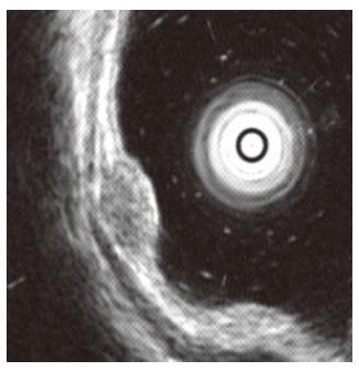
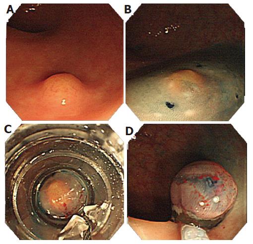
Fifteen patients diagnosed as rectal carcinoid tumor (less than 10 mm) in our hospital from 1993 to 2002 were enrolled in this study. No patients had symptoms of carcinoid syndrome. Examination with a high-frequency ultrasonographic probe (EU-M20; 20MHz, Olympus, Tokyo, Japan) inserted through the biopsy channel of the endoscope was performed for evaluation of carcinoid tumors remained in submucosal layer (Figure 1). After informed consent was obtained, these patients were randomly assigned into two groups according to a table of random permutations. Group 1 (n = 7) carcinoid tumors were treated with endoscopic mucosal resection using a snare with a conventional single-channel colonoscopy. Briefly, after injection of submucosal saline, the lesion was simply resected with snaring. Group 2 (n = 8) carcinoid tumors were treated by endoscopic resection using a ligation device (varioligator kit; TOP Co. Ltd, Tokyo, Japan), with modification of the method used in gastric carcinoma[4,5]. A ligation device attached to a conventional single-channel colonoscopy (PCF200, PCF240 Olympus, Co., Tokyo, Japan), and endoscopic resection using a ligation device was performed as follows: (1) submucosal saline solution was injected beneath the tumor to elevate it for reducing a risk of perforation and resection margin involvement; (2) the carcinoid tumor was aspirated into the ligation device; (3) snare resection was performed below the band by using blend electrosurgical current (Figure 2). This study was performed according to the guidelines of the Committee on Clinical Practice of our hospital.
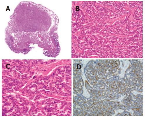
All resected specimens were examined microscopically for histopathological diagnosis, cut (lateral) and deep (vertical) margin involvement, and vascular and/or lymphatic invasion (Figure 3). Completeness of resection was evaluated by the histopathological examination as absence of carcinoid in the margin of the resected specimen.
Tested groups were evaluated using chi-square test or student’s t test. P values of 0.05 or less were considered statistically significant.
The results are shown in Table 1. Average age of group 1 patients was 60.2 years (range: 34-75 years), and average age of group 2 was 62.6 years (range: 51-77 years). Female/male ratios were 3:4 in group 1 and 3:5 in group 2. Thirteen tumors were located in the lower and 2 in the upper portion of the rectum, as 9 sessile and 6 semipedunculated small protrusions of the mucosa. The size of the tumors varied from 3 mm to 9 mm with mean diameter of 6.3 mm in group 1, and from 4 mm to 10 mm with mean diameter of 6.2 mm in group 2. Neither vascular nor lymphatic invasion was observed in any resected carcinoid tumor. Background characteristics of patients were not different between groups 1 and 2.
| Age (yr) | Sex | Size (mm) | Margin | Additional therapy |
| Group 1 | ||||
| 74 | M | 3 | Negative | (-) |
| 34 | F | 5 | Negative | (-) |
| 61 | M | 7 | Negative | (-) |
| 46 | F | 8 | Positive | Transanal resection |
| 64 | M | 6 | Negative | (-) |
| 75 | M | 6 | Positive | Ligation |
| Group 2 | ||||
| 55 | M | 5 | Negative | (-) |
| 77 | M | 9 | Negative | (-) |
| 70 | M | 7 | Negative | (-) |
| 51 | F | 4 | Negative | (-) |
| 63 | M | 10 | Negative | (-) |
| 74 | F | 4 | Negative | (-) |
| 60 | F | 5 | Negative | (-) |
| 51 | M | 6 | Negative | (-) |
Complete resection rate was compared in two groups with histopathological examination. The rate of complete removal of carcinoid tumor in group 1 with simple snaring resection was 42.9% (3/7), which was significantly lower compared to that in group 2 with resection using a ligation device (100%, 8/8; P = 0.024). The three patients had tumor involvement of deep margin without lateral resection margin. These patients of carcinoid tumor in group 1 were treated with additional treatment: one patient was treated by trans-anal resection and two patients were resected with a ligation device. All patients were followed up more than 3 years, and recurrence of carcinoid tumors was not observed in any patient enrolled in this study.
Rectum is the most common part of carcinoid tumors in the gastro-intestinal tract in Japan[7]. Prevalence of rectal carcinoid has been reported as 0.07% (16/21 522) in healthy subjects[3] and 0.10% (8/7 665) in subjects of total colonoscopy in Japan[8]. Most of rectal carcinoid tumors are less than 10 mm in size, and these tumors remain in the rectum without metastasis and can be treated by local resection[1,2]. These carcinoid tumors are mainly located in the lower part of rectum as shown in this study[3,8-10].
Rectal carcinoid tumors are treated by several methods including endoscopic resection[11-13] and surgical operation with trans-anal approach[14]. Carcinod tumors less than 10 mm can be treated by local resection, but most of carcinoid tumors extend into the submucosa, and polypectomy can not remove these tumors completely as shown in the previous study[4]. Complete resection of submucosal tumors requires other method than simple polypectomy. Endoscopic mucosal resection following submucosal injection of saline solution should be useful for the resection.
This randomized control study compared endoscopic resection with a ligation device to conventional endoscopic resection with a snare just after saline injection. As a result, rectal carcinoid tumors less than 10 mm could be removed completely by endoscopic resection with a ligation device without any procedure-related complications. This result confirmed the previous historical control trial performed by Ono et al[4] who showed usefulness of endoscopic resection with a ligation device but their follow-up period was limited (median follow-up period: 10.5 mo). In this study, we followed up all patients for 3 years without any recurrence.
In conclusion, endoscopic resection with a ligation device might be a most applicable procedure for rectal carcinoid tumors less than 10 mm remained within submucosal layer, because this procedure is simple, minimally invasive, and safe for resection.
S- Editor Wang J L- Editor Kumar M E- Editor Liu Y
| 1. | Koura AN, Giacco GG, Curley SA, Skibber JM, Feig BW, Ellis LM. Carcinoid tumors of the rectum: effect of size, histopathology, and surgical treatment on metastasis free survival. Cancer. 1997;79:1294-1298. [PubMed] [DOI] [Cited in This Article: ] [Cited by in F6Publishing: 3] [Reference Citation Analysis (0)] |
| 2. | Soga J. Carcinoids of the rectum: an evaluation of 1271 reported cases. Surg Today. 1997;27:112-119. [PubMed] [DOI] [Cited in This Article: ] [Cited by in Crossref: 129] [Cited by in F6Publishing: 110] [Article Influence: 4.1] [Reference Citation Analysis (0)] |
| 3. | Matsui K, Iwase T, Kitagawa M. Small, polypoid-appearing carcinoid tumors of the rectum: clinicopathologic study of 16 cases and effectiveness of endoscopic treatment. Am J Gastroenterol. 1993;88:1949-1953. [PubMed] [Cited in This Article: ] |
| 4. | Ono A, Fujii T, Saito Y, Matsuda T, Lee DT, Gotoda T, Saito D. Endoscopic submucosal resection of rectal carcinoid tumors with a ligation device. Gastrointest Endosc. 2003;57:583-587. [PubMed] [DOI] [Cited in This Article: ] [Cited by in Crossref: 105] [Cited by in F6Publishing: 104] [Article Influence: 5.0] [Reference Citation Analysis (0)] |
| 5. | Suzuki Y, Hiraishi H, Kanke K, Watanabe H, Ueno N, Ishida M, Masuyama H, Terano A. Treatment of gastric tumors by endoscopic mucosal resection with a ligating device. Gastrointest Endosc. 1999;49:192-199. [PubMed] [DOI] [Cited in This Article: ] [Cited by in Crossref: 67] [Cited by in F6Publishing: 66] [Article Influence: 2.6] [Reference Citation Analysis (0)] |
| 6. | Sakata H, Iwakiri R, Okamoto K, Watanabe K, Matsunaga K, Oda K, Ootani H, Shimoda R, Tsunada S, Mizuguchi M. Effective endoscopic resection using a ligation device for rectal carcinoid tumor. Gastroinetest Endosc. 2002;55:AB118. [Cited in This Article: ] |
| 7. | Soga J. Carcinoid tumors: A statistical analysis of a Japanese series of 3,126 reported and 1,180 autopsy cases. Acta Med Biol. 1994;42:87-102. [Cited in This Article: ] |
| 8. | Koyama N, Yoshida H, Nihei M, Sakonji M, Wachi E. Endoscopic resection of rectal carcinods using double snare polypectomy technique. Dig Endosc. 1998;10:42-45. [DOI] [Cited in This Article: ] [Cited by in Crossref: 8] [Cited by in F6Publishing: 8] [Article Influence: 0.5] [Reference Citation Analysis (0)] |
| 9. | CALDAROLA VT, JACKMAN RJ, MOERTEL CG, DOCKERTY MB. CARCINOID TUMORS OF THE RECTUM. Am J Surg. 1964;107:844-849. [PubMed] [DOI] [Cited in This Article: ] [Cited by in Crossref: 76] [Cited by in F6Publishing: 81] [Article Influence: 1.4] [Reference Citation Analysis (0)] |
| 10. | Grablowsky OM, Ribaudo JM, Ray JE. Carcinoid tumors of the rectum: Experience with 38 cases. Dis Colon Rectum. 1974;17:532-535. [PubMed] [DOI] [Cited in This Article: ] [Cited by in Crossref: 11] [Cited by in F6Publishing: 11] [Article Influence: 0.2] [Reference Citation Analysis (0)] |
| 11. | Kajiyama T, Hajiro K, Sakai M, Inoue K, Konishi Y, Takakuwa H, Ueda S, Okuma M. Endoscopic resection of gastrointestinal submucosal lesions: a comparison between strip biopsy and aspiration lumpectomy. Gastrointest Endosc. 1996;44:404-410. [PubMed] [DOI] [Cited in This Article: ] [Cited by in Crossref: 46] [Cited by in F6Publishing: 41] [Article Influence: 1.5] [Reference Citation Analysis (0)] |
| 12. | Berkelhammer C, Jasper I, Kirvaitis E, Schreiber S, Hamilton J, Walloch J. "Band-snare" resection of small rectal carcinoid tumors. Gastrointest Endosc. 1999;50:582-585. [PubMed] [DOI] [Cited in This Article: ] [Cited by in Crossref: 30] [Cited by in F6Publishing: 31] [Article Influence: 1.2] [Reference Citation Analysis (0)] |
| 13. | Iishi H, Tatsuta M, Yano H, Narahara H, Iseki K, Ishiguro S. More effective endoscopic resection with a two-channel colonoscope for carcinoid tumors of the rectum. Dis Colon Rectum. 1996;39:1438-1439. [PubMed] [DOI] [Cited in This Article: ] [Cited by in Crossref: 35] [Cited by in F6Publishing: 36] [Article Influence: 1.3] [Reference Citation Analysis (0)] |
| 14. | Araki Y, Isomoto H, Shirouzu K. Clinical efficacy of video-assisted gasless transanal endoscopic microsurgery (TEM) for rectal carcinoid tumor. Surg Endosc. 2001;15:402-404. [PubMed] [DOI] [Cited in This Article: ] [Cited by in Crossref: 14] [Cited by in F6Publishing: 16] [Article Influence: 0.7] [Reference Citation Analysis (0)] |


