Copyright
©2006 Baishideng Publishing Group Co.
World J Gastroenterol. May 14, 2006; 12(18): 2858-2863
Published online May 14, 2006. doi: 10.3748/wjg.v12.i18.2858
Published online May 14, 2006. doi: 10.3748/wjg.v12.i18.2858
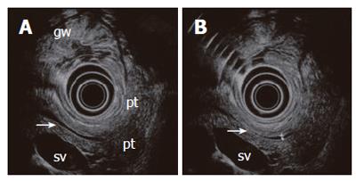
Figure 5 A normal pancreas imaged from the stomach with a 6 MHz echoendoscope.
The compressed posterior gastric wall is closely related to the surface of the pancreas. The pancreatic duct is seen (arrow) with minor (A) and slightly increased transducer pressure (B). The non-compressed gastric wall with folds (gw) is seen on the opposite side of the transducer. Pancreatic tail (pt), splenic vein (sv).
- Citation: Odegaard S, Nesje LB, Hoff DAL, Gilja OH, Gregersen H. Morphology and motor function of the gastrointestinal tract examined with endosonography. World J Gastroenterol 2006; 12(18): 2858-2863
- URL: https://www.wjgnet.com/1007-9327/full/v12/i18/2858.htm
- DOI: https://dx.doi.org/10.3748/wjg.v12.i18.2858


